Sylvian fissure (brain): what is it, functions and anatomy?

It is one of the most visible clefts on the surface of the brain. We explain its characteristics.
Our brain is one of the most important and complex organs of our body.It is full of different structures, areas and regions of great importance that govern different basic aspects for the maintenance of life.
These structures require a space to exist, a space that is limited by the bony structure that protects the organ: the skull. And some of these structures could be really big, as is the case with the cerebral cortex. Fortunately, throughout our development, the brain becomes more compact, the cerebral cortex growing in such a way that it forms different folds (which gives the brain its characteristic appearance). And with these folds also appear the furrows between them. One of the most famous is the lateral sulcus or Sylvian fissure..
Fissures and grooves
Before going into detail about what is the Sylvian fissure, we must stop for a moment and first ask ourselves how our brain is structured. In this way we will better understand the path that this fissure traces along the cerebral cortex.
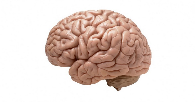
Seen from the outside, the brain appears as a relatively compact a relatively compact mass, the cerebral cortex being full of folds, so that the whole of it is so that the whole of it fits inside the skull. The existence of these folds also generates the existence of different clefts, which are called fissures or grooves. The concave parts, the ones that protrude, are the gyri or convolutions.
Thus, a cerebral sulcus or fissure is considered to be the one that a cleft or gap left by the cerebral cortex as it folds back on itself during development, and which, when viewed from the surface, gives an idea of the boundaries of the lobes of the brain. and that, seen from the surface, gives an idea of what are the limits of the lobes of the brain.
The Sylvian fissure: what is it and what areas does it separate?
The Sylvian fissure or lateral sulcus is, together with the Rolando's fissure, one of the most visible and recognizable fissures or sulci in the human brain. It is located in the lower part of the two cerebral hemispheres and then runs transversely through a large part of the brain. This furrow appears horizontally, being located in the naso-lambdoid line.
It is one of the most relevant sulci, since it separates the temporal and temporal lobes. separates the temporal and parietal lobes and in its lower part the frontal and temporal lobes.. This is the deepest cleft in the entire brain, to the point that in its depths hides the so-called fifth cerebral lobe: the insula. It also contains the transverse temporal gyrus, which is involved in the auditory system.
It should also be noted that the middle cerebral artery the middle cerebral artery, also known as the sylvian artery, passes through it. artery, which irrigates the different cerebral regions of the area.
This fissure is one of the first to appear throughout our development, being already visible in fetal development. Specifically, it can often be observed from the fourteenth week of gestation. Their morphology and depth will evolve as the fetus develops.
Branches
The cisura of Sylvius can be divided into several branchesThe name of these branches gives an idea about their orientation. The name of these gives an idea about their orientation.
Between the first and the second the third frontal gyrus, and specifically the pars triangularis (corresponding to Brodmann's area). (corresponding to Brodmann's area 45). In the horizontal branch the pars orbitalis (area 47) and the pars opercularis (corresponding to area 44) between the oblique and vertical trifurcation branches. These areas are associated with language production.
Diseases and disorders with alterations in this cisura
The Sylvian fissure is a groove that all or practically all human beings possess. However, there are diseases in which this fissure is not correctly formed or is altered for some reason. Among them we can find examples in the following pathologies.
Alzheimer's disease and other dementias
Patients with Alzheimer's disease often present during the course of their disease with an enlargement of the fissure of SylviusThis enlargement is the result of neuronal tissue degeneration. This anomaly can also be found in other dementias and neurodegenerative diseases, which with the passage of time kill nerve cells and cause the brain to have a withered appearance, with large grooves and very pronounced folds. This means that its effects are not limited to the sylvian fissure, but are felt throughout the cortex in general.
2. The absence of cerebral sulci: lissencephaly
Lissencephaly is an anomaly generated during neurodevelopment in which the brain appears smooth and either without or with few gyri and cysplasias, an alteration caused by a deficit or absence of neuronal migration or by an excess of neuronal migration.. This phenomenon may have genetic causes or be due to alterations produced during embryonic development.
It can present in two ways: the complete, also called agyria, in which neither gyri or cerebral furrows are developed, and the incomplete or pachygyria in which there are some, although they are few and very wide. There is usually a deficient covering of brain parenchyma in the Sylvian fissure.
In general, the prognosis is not good, and the disease is associated with a short life expectancy, presenting symptoms such as seizures, Respiratory problems and intellectual disability, although in some cases there are no major problems.
3. Opercular syndrome
The opercular or perisylvian syndromeThe syndrome, in which there are motor control problems or even paralysis in the face area, is also linked to the Sylvian fissure because there are problems in the operculum, the brain areas surrounding the Sylvian fissure and corresponding to the part not directly visible from the outside.
4. Cerebrovascular alterations
The middle cerebral artery passes through the Sylvian fissure. That is why alterations in this area can also affect this part of the circulatory system, which is capable of generating problems such as aneurysms, hemorrhages or embolisms.
Bibliographic references:
- Chi J.G.;, Dooling, E.C. & Gilles, F.H. (January 1977). "Gyral development of the human brain". Annals of Neurology 1 (1): 86-93.
- Kandel, E.R.; Schwartz, J.H.; Jessell, T.M. (2001). Principles of Neuroscience. Madrid: MacGrawHill.
- Santos, L. (2000). Synthesis of human anatomy. Conceptual keys and Atlas of basic schemes. Ediciones Universidad de Salamanca.
(Updated at Apr 13 / 2024)