Magnetoencephalography: what is it and what is it used for?

A summary of the characteristics, advantages and disadvantages of magnetoencephalography.
Magnetoencephalography is one of the best-known neuroimaging techniques used both in clinical intervention programs and in research on the human brain. It is therefore an example of how technology helps us to know ourselves better.
In this article we will see what magnetoencephalography consists of and how it works, and what its uses are.and what are its uses.
Understanding the brain from new technologies
There is no doubt that the brain is a system made up of millions of highly complex biological processes, among which language is one of the most important.Among these are language, perception, cognition and motor control. That is why for thousands of years this organ has attracted a great deal of interest from all kinds of scholars who have provided various hypotheses about its functions.
Some years ago, in order to measure cognitive processes, behavioral measurement techniques were used; for example, reaction time measurements and pencil and paper tests. Later, during the 1990s, technological breakthroughs made it possible to record brain activity related to these cognitive processes. This was a great qualitative leap in this area of research and a complement to the traditional techniques that are still in use today.
Thanks to these advances, it is now known that billions of billions of brain function involves trillions of neurons that are interconnected with each other.These connections are set in motion by the brain's electrical impulses.
Each neuron can be said to work as if it were a "small electrochemical pump" containing ions, which are electrically charged, and are in continuous movement both inside and outside the cell membrane of the neuron. When the neurons are charged, they provide a flow of current into the cells, and these in turn are stimulated, causing what is known as "neuron firing".causing what is known as an action potential that causes the neuron to trigger the flow of charged ions.
This electrical potential travels until it reaches the presynaptic region and then releases neurotransmitters into the synaptic space that access the postsynaptic cell membrane and immediately cause changes in intra- and extracellular ionic flux.
At the moment when several neurons and synaptically interconnected cells are activated simultaneously, they provide an electric current flow accompanied by a magnetic field. a flow of electrical current accompanied by a magnetic field and, accordingly and, accordingly, flow towards the cerebral cortex.
It is estimated that in order to create a magnetic field, measurable by means of measuring instruments placed on the head, it is necessary for 50,000 neurons to be activated simultaneously, it is necessary that 50,000 neurons or more are active and interconnected.. If it were the case that there were electric currents moving in opposite directions, the magnetic fields accompanying each current would cancel each other out (Hari and Salmelin, 2012; Zhang et al., 2014).
These complex processes can be visualized thanks to neuroimaging techniques, including one that we would like to highlight and will address in more detail in this article, magnetoencephalography.
What is magnetoencephalography?
Magnetoencephalography (MEG) is a neuroimaging technique used to a neuroimaging technique used to measure the magnetic fields produced by electrical currents in the brain.. These electrical currents are produced by neural connections throughout the brain in order to produce multiple functions. Each function produces certain brain waves and this would allow us to detect, for example, whether a person is awake or asleep.
The MAG is also a non-invasive medical test; therefore, during its use, no instrument needs to be introduced into the skull to detect the interneuronal electrical signals. This tool makes it possible to study the human brain 'in vivo', so that we can detect various mechanisms of the brain in full operation while the person receives certain stimuli or performs some activity.. At the same time it allows us to locate any anomaly if any (Del Abril, 2009).
With MEG we can visualize moving three-dimensional images with which we can accurately detect, in addition to anomalies, their structure and function. This allows professionals to investigate whether there is any relationship with the personality of the subjects who present these anomalies, to study whether genetics plays a relevant role, and even to contrast whether they influence cognition and emotions.
Who is in charge and where is MEG usually used?
The specialized professional who is in charge of performing these brain evaluation tests is the medical radiologist.
This test, as well as all other neuroimaging techniques, is usually performed in hospital settings where all the necessary equipment is available.
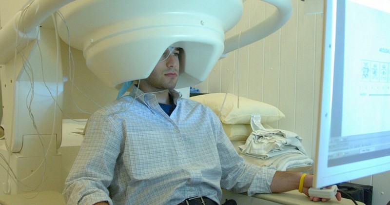
The systems that perform MEG are carried out in a specialized room that must be shielded in order to prevent interference that could be produced by the strong magnetic signals that would be produced by the environment if it were performed in any other place.
To carry out this test The patient is seated and a "helmet" containing magnetic sensors is placed over the head.. A computer detects the signals that provide the MEG measurement.
Other techniques that make it possible to study the brain 'in vivo'.
Neuroimaging techniques, also known as neuroradiology tests, make it possible to obtain an image of the brain structure in full operation. These techniques allow the study of disorders or anomalies of the central nervous system in order to find a treatment for them..
According to Del Abril et al. (2009) the most commonly used techniques in recent years, apart from magnetoencephalography, are the following.
1. Computerized axial tomography (CT)
This technique is used through a computer that is connected to an X-ray machine.. The objective is to capture a series of detailed images of the interior of the brain, taken from different angles.
2. Magnetic Resonance Imaging (MRI)
This technique uses a large electromagnet, radio waves and a computer to capture detailed images of the brain. With MRI, images of higher quality are obtained than those obtained with CT.. This technique was a great advance in brain imaging research.
3. Positron Emission Tomography (PET)
It is considered one of the most invasive techniques. It is used to measure the metabolic activity of different regions of the brain.
This is achieved by injecting the patient with a radioactive substance that binds to glucose and then binds to the membranes of the cells of the central nervous system through the bloodstream. the central nervous system through the bloodstream.
The glucose accumulates at a high rate in areas of increased metabolic activity. This makes it possible to identify a decrease in the number of neurons in a certain area of the brain, if hypometabolism is detected.
4. Functional magnetic resonance imaging (fMRI)
This technique is another variant used to visualize the regions of the brain that are active at certain times or when performing some activity; this is achieved by detecting the increase of oxygen in the Blood in these more active areas. It allows to obtain images with a better resolution than other functional imaging techniques..
5. Electroencephalogram (EEG)
A technique initiated in the 1920s that is used to measure the electrical activity of the brain by placing electrodes on the skull.
The purpose of this tool is to to investigate the brain wave patterns associated with specific behavioral states (e.g., brain waves, brain waves, etc.). (e.g. beta waves are associated with alertness and wakefulness, while delta waves are associated with sleep) and also to detect possible neurological disorders (e.g. epilepsy).
A great advantage that MEG has over EEG is the ability to reveal the three-dimensional location of the group of neurons that is generating the magnetic field being measured.
Advantages and disadvantages of magnetoencephalography
As with any resource to make the brain a comprehensible reality and capable of providing relevant data, magnetoencephalography has certain advantages and disadvantages. Let us see what they are.
Advantages
According to with Zhang, Zhang, Reynoso and Silva-Pereya (2014) among the advantages presented by this revolutionary brain measurement technique the following stand out.
As previously mentioned, it is a noninvasive test, so that it is not necessary to penetrate the interior of the skull with any type of instrumentation. The technique is specialized to measure the magnetic fields emitted by neuronal currents in the various regions of the brain. Moreover, it is the only completely non-invasive neuroimaging technique. Of course, it is painless to use.
In addition, it allows the possibility of to see functional images of the brain at times when it is deduced that there might be a disorder but there is no anatomical evidence but there is no anatomical evidence to prove it. That is why this test shows the local point of brain activity with high accuracy.
Another advantage that has been found is that it also offers the possibility of to test infants who have not yet acquired the ability to emit behavioral responses..
Finally, according to Maestu et al. (2005) the MEG signal does not degrade as it passes through the different tissues, which is the case with MEG currents.which is the case with EEG currents. This allows magnetoencephalography to measure neuronal signals directly and in a matter of milliseconds.
Disadvantages
According to Maestu et al. (2005), MEG presents some limitations that prevent it from being the definitive technique in the field of the study of cognition.. These limitations are:
- Impossibility of capturing sources deep in the brain.
- High sensitivity to the environment in which the test is performed.
(Updated at Apr 12 / 2024)