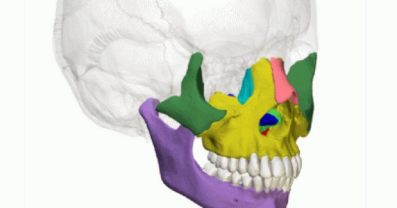
Bones of the face: types, characteristics and location.

A summary classification of the different bones of the face, described.
We see it every day in the mirror and, although we easily recognize it as our own, many of us know little or nothing of what lies beneath the skin.
The face is a familiar part of us all, almost the most familiar. It is what gives us, so to speak, our outward personality, for in a world where appearances matter, the face is of the utmost importance: it is our calling card.
Underneath it we can find many bones, many of them unknown to most people since, despite being so important, it is the anatomical part that is least studied in schools. For this reason we bring here the list with the main bones of the facewhat structures they form and what they are inserted with.
What are the bones of the face?
Although we are not very narcissistic people, the face is that part of the body that worries us the most, since our appearance depends a lot on it. But although it is that part of the body that we see every day, looking at us in the mirror while we get ready in the morning, it is also that great unknown since the layers of skin on it prevent us from seeing the complexity of its bones.
Anatomically, we can define the face as a bony conglomerate located in the lower and anterior part of the head.. This structure is composed of many bones in spite of being a relatively small region, being the total of bony structures that can be found in it about fourteen. Of these fourteen bones, six are pairs and two are odd or single, located near the midline of the face and housing in their various cavities the organs of most of the senses.
1. Upper jaw
The upper jaw is composed of a pair of short, irregular bones flattened from the inside out.. It has two faces, one internal and one external, as well as four edges and four angles. Its lower edge serves as an insertion for the teeth of the upper arch, i.e., the teeth of the upper jaw.
This structure articulates with:
- The maxilla of the opposite side in the midline.
- The frontal and ethmoid, together with the bones proper to the nose above.
- The palatines and the vomer towards the middle and behind.
- It shapes part of the ocular orbit and nostrils.
2. Palatines
The palatines are a pair of short and irregular bones, one on the right side and the other on the left side. They are located behind the maxilla with which they articulate forward..
These bones articulate with:
- The other palatine on the opposite side.
- The sphenoid bone behind.
- The vomer and inferior nasal conchae above.
- They form part of the nostrils.
3. Zygomatics or malar bone
The zygomatics are two short, irregular bones located in the outermost part of the face, just at the level of the cheeks, and are in fact also known as the zygomatic or malar bone. and, in fact, are also known as the malar bones or cheekbones. Their shape is flattened from outside to inside and, having four edges with their respective four angles, their shape suggests that of a quadrilateral. It has two faces, one external and one internal, which are located on the inferior and lateral face to the frontal.
The zygomatics articulate with:
- The frontal from above.
- The upper jaws underneath.
- The temporals on the sides.
- They shape part of the ocular orbit.
4. Nasal bone
The nasal bone, also called the nasal bone, is a paired bone placed on each side of the midline and located just at the top of the human nose, being in fact the only external structure of that region that is located just above the nose.It is in fact the only external structure of that region that is composed of bony tissue, namely a quadrilateral lamina with two faces and four edges.
This structure articulates with:
- The frontal bone above.
- The upper jawbone below.
- The other side of the nose and the ethmoid bone.
- It forms part of the nostrils.
5. Lower turbinates or nasal conchae
The turbinates are two bones located in the lower part of the nostrils.. Their other name, inferior nasal conchae, indicates that they are part of the nostrils. They have two faces, one internal and one external, two edges and two extremities.
The turbinates of the face articulate with:
- The ethmoid and maxilla from above.
- The palatine behind.
- The lacrimals in front.
6. Unguis or lacrimal bones
The lacrimal bones are a paired bone located in the anterior part of the internal face of the fossa that forms the ocular orbit. They are also characterized by the fact that they contribute to the formation of the nostrils and constitute a small bony lamina.. Its shape is quadrilateral and irregular, having two faces and four edges.
7. Vomer
The vomer is a bone with a curious name that happens to be unique and odd, unlike the it is unique and odd, unlike most of the bones that make up the face.. It is located in the facial midline, constituting the posterior part of the nasal septum. It is a very thin quadrilateral lamina with two faces and two edges.
The vomer articulates with:
- The ethmoid and sphenoid above.
- The upper maxillae and palatines below.
- It is part of the nasal septum.
8. Lower maxilla or mandible
The lower jaw is a large, single, irregularly shaped but symmetrical bone that is located in the center of the facial midline, although in its lower part.The lower jaw is a large, single, irregularly shaped but symmetrical bone. It has a horseshoe shape and is joined to other bones by a mobile joint, which gives it a certain freedom of movement.
It is thanks to this joint that we can move the lower jaw to be able to chew, speak or gesture. It has two faces, one anterior and one posterior, two lateral extremities or ascending branches and two edges, an upper one that gives insertion to the teeth of the lower arch.
Bone attachments of the face
Now that we have seen the 8 types of bones of the face, which actually constitute the 14 bones of this anatomical region, let's talk about the bony unions they form. Four main structures arise from the union of the bones of the face: the ocular orbit, the nasal fossae, the pterygomaxillary fossa and the palatine vault.
1. Ocular orbit
The ocular orbits are excavated cavities that are widely recognized as the hollows where the eyes are located.. These cavities are located between the face and the rest of the skull and are characterized by being located on both sides of the face, one on the right and the other on the left, presenting a quadrangular pyramid shape with an anterior base.
Inside the orbit we can see four walls:
- Superior or roof: it is formed by the horizontal portion of the frontal and the lesser wing of the sphenoid.
- Inferior or floor: formed by the pyramidal process of the maxilla, the orbital process of the zygomatic and the orbital process of the palatine.
- Internal: formed by the ascending process of the maxilla, the lacrimals and the orbital plate of the ethmoid.
- External: formed by the greater wing of the sphenoid and the orbital processes of the zygomatic and frontal bone.
2. Nostrils
We can describe the nostrils as long flattened corridors, which are characterized by being transversely located to the right and left of the midline.. Each nostril has four walls and two openings, one anterior and one posterior. Delving into these four walls, we observe:
- Outer wall: formed by six bones, which are the maxilla, sphenoid, palatine, lacrimal, inferior nasal conchae and ethmoid.
- Inner wall: it is constituted by the nasal septum, which in turn is formed by the vomer and the perpendicular lamina of the ethmoid.
- Upper wall or roof: it is formed by the nasal bones, the nasal spine of the frontal bone, the horizontal lamina of the ethmoid and the body of the sphenoid.
- Inferior wall or floor: it is formed by the palatine process of the upper jaw and the horizontal lamina of the bone.
3. Pterygomaxillary fossa
The pterygomaxillary fossa is a small region located inside the zygomatic fossa.. This structure is shaped like a quadrangular pyramid with four walls, a base and an apex.
4. Palatal vault
The palatine vault is a horseshoe-shaped region bounded behind the posterior border of the palatine. In front and on the sides is the alveolar border of the maxilla.
(Updated at Apr 14 / 2024)