Corona radiata: characteristics and functions of this part of the brain.
Let's see what the corona radiata is and what functions it has in the human brain.
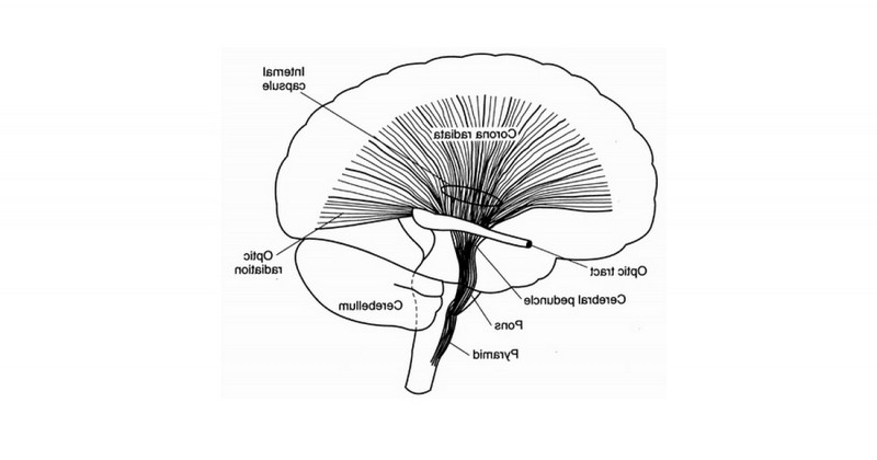
The corona radiata is a brain structureIt is made up of nerve fibers that project into the brain to form the internal capsule, a structure that connects to the cortex.
Located in both hemispheres, each corona radiata connects with its opposite through the corpus callosum.
Below we explain in more detail what this brain structure consists of, what are its characteristics, structure and functions.
Corona radiata: what is it?
The corona radiata or corona radiata is a brain structure formed by nerve fibers (nerve substance). formed by nerve fibers (white matter) that form the internal capsule of the brain.The cerebral cortex, a region that connects the cerebral cortex with lower areas of the brain and the spinal cord.
This region of the brain is called the corona radiata because its nerve fibers project in a kind of corona-like structure.
The nerves of the corona radiata transport information between the brain cells of the cerebral cortex and the cells in the brainstem.. The cortex is the area responsible for processing conscious information, while the brainstem is responsible for the connections between the spinal cord and the brain. Both are involved in sensation and motor function, and the corona radiata connects the motor and sensory nerve pathways between these structures.
The corona radiata may be affected by diseases capable of affecting the cerebral white matter.The white matter of the brain, such as multiple sclerosis, causes important intellectual, social and affective dysfunctions.
Structure and composition.
The corona radiata is composed of a large set of projection fibers; a group of afferent fibers, which transmit information to the cerebral cortex; and a group of efferent fibers, which travel in the reverse direction, handling information from the cortex.
In each cerebral hemisphere and underlying the cortex, there is a large amount of white matter formed by fibers, which can be formed by fibers, which can be: association fibers, in charge of connecting different parts within the same cerebral hemisphere; commissural fibers, which connect regions between the two hemispheres; and projection fibers, which connect the cerebral cortex with distant underlying structures.
In the brain, projection fibers are bundled within the internal capsule. This structure is a compact band of white matter composed of ascending and descending nerve fibers that connect the cerebral cortex with the brainstem and spinal cord.
The projection fibers of the internal capsule fan out to form the corona radiata. Many of these fibers establish reciprocal connections between the thalamus and the cerebral cortex..
These connections configure the following structures: the anterior thalamic radiation, formed by fibers connecting the dorsomedial nucleus of the thalamus and the prefrontal cortex; the medial thalamic radiation, which includes the somatosensory projection from the thalamus to the parietal lobe; the posterior thalamic radiation, connecting the thalamus and the occipital lobe cortex; and the inferior thalamic radiation, formed by fibers connecting the thalamic nuclei with the temporal lobe cortex, forming the auditory radiation.
Main functions
As we have seen, the nerve fibers of the corona radiata converge to form the internal capsule. This, in turn, divides another structure called the striatum or striatal nucleus, which receives information from the temporal lobe cortex.which receives information from the cerebral cortex and forms part of the basal ganglia.
The function of the basal ganglia is to regulate and control movements, manage learning related to automated procedures (e.g., driving a vehicle), intervene in motivational and emotional processes, or manage activities related to planning.
The internal capsule is directly related to two of the structures that make up the basal ganglia: the caudate nucleus and the putamen. These two regions are separated by the descending fibers of the internal capsule.
The caudate nucleus participates in the modulation of movement in an indirect way; and the putamen is mainly responsible for the motor control of the body and plays a relevant role in operant conditioning.
Lesions affecting this brain structure
The corona radiata can be damaged by various causes, such as stroke. The effusions involve small branches of Blood vessels and those that affect the corona radiata, are usually called subcortical, lacunar or white matter effusions.are generally referred to as subcortical, lacunar or white matter effusions.
The reason why this region is called white matter is because it is highly myelinated, which means that it is protected by a special type of fatty tissue that insulates and helps nerve cells: myelin. They are also called subcortical strokes because they are found in the subcortical and deeper region of the brain, in contrast to cortical or more superficial regions.
People who suffer accidents or damage to an area such as the corona radiata suffer from what is termed as cerebrovascular disease, which is characterized by narrowed blood vessels that are prone to develop clots. and prone to develop blood clots in the brain.
Occasionally, strokes involving the corona radiata may be relatively small and cause no symptoms. In such a case, they are called silent strokes.
On the other hand, a stroke in a region such as the corona radiata may produce non-specific symptoms, such as loss of autonomy and daily living skills, a predictor of stroke.A stroke is a predictor of stroke, even when there are no significant signs on a brain MRI or CT scan.
In addition to stroke, there are other causes of damage to the corona radiata, such as: brain tumors, metastatic spread of cancer, cerebral hemorrhage, head trauma or brain infections.
However, there are two keys to prevention, there are two keys to prevention: healthy lifestyle habits and regular medical care.. Not smoking, eating a healthy diet, relaxing and avoiding stressful situations or dealing with medical problems such as high cholesterol or hypertension are some of the protective factors that will help us to prevent diseases and strokes.
Bibliographic references:
- Gutman, D.H., Scherer, S. (1989). Magnetic resonance imaging of ataxic hemiparesis localized to the corona radiata. Stroke. 1989;20:1571-1573.
- Richard, S.S (2007). Clinical neuroanatomy. Médica Panamericana.
- Sage, J., Lepore, F.E. (1983). Ataxic hemiparesis from lesions of the corona radiata. Arch Neurol; 40:449-450.
(Updated at Apr 13 / 2024)