Epiblast: what is it and what are its characteristics?

The epiblast is one of the main cell layers of embryos; let's see what it looks like.
Embryology is a subdiscipline of genetics and biology that studies morphogenesis, embryonic and nervous development from gametogenesis to the moment of birth of living beings. Human life begins with an ovum and a spermatozoon, two specialized haploid cells (n) which, after sexual intercourse, unite and form a zygote (2n).
Humans have 23 pairs of chromosomes in the nucleus of almost all our cells, i.e. a total of 46. At the moment of fertilization, the two haploid cells mentioned above fuse, so half of the genetic information that codes us comes from our father and the other half from the mother. This simple mechanism explains the keys to inheritance in our species and in many other living beings, since genetic recombination processes and spontaneous mutations occur that generate variability in living beings in the long term.
Beyond the genetic mechanism of reproduction and the formation of a viable embryo, it is really interesting to know how we go from being a fusion of two cells to a fetus, with differentiated and clear anatomical structures. Today we tell you all about the epiblast, one of the cell lineages present during gastrulation of embryonic development in mammals, reptiles and birds.
What is the epiblast?
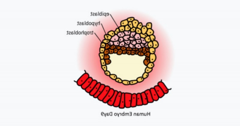
In the field of embryology, an epiblast can be defined as a layer of embryonic cells that appears during gastrulation (together with the hypoblast) and gives rise to the mesoderm and ectoderm.. The functionality of this cell lineage can be intuited if we turn to its etymological basis: epi- means envelope, while the Greek term βλαστός refers to a germ, bud or sprout. In the epiblast resides the germ of life, for without it human development could not be completed.
At the histological level, this layer of cells is described as a columnar epithelium rich in microvilli in its apical portion.. These appear around day 8 after fertilization, and undergo epithelial-mesenchymal change throughout development to give rise to the precursor layers of the various organs and structures of living beings.
We have introduced many complex terms out of the blue, but don't worry. To start from zero and to understand the definition provided, we dissect each of the complex words exposed in the following lines.
What is gastrulation?
Gastrulation is one of the stages of early embryonic development following implantation of the blastocyst in the endometrium.. After implantation of the product of the female egg and the male sperm, between the 4th and 5th week of pregnancy, the embryo begins to undergo very important changes, among which are the processes described in the following lines.
It should be noted that the first cell body of interest that we encounter during gestation is the blastocyst, already mentioned. This is composed of about 200 cells and appears the first 5-6 days after fertilization.
It is the stage of development prior to the implantation of the embryo in the maternal uterus, and is differentiated into 2 main structures: the inner cell mass (ICM) or embryoblast, which will subsequently form the embryo, and the trophoblast, the outermost cell layer that protects the blastocyst.
Gastrulation is a process by which a trilaminar embryo is formed by the migration of cell populations located in the epiblast.. These laminae correspond to the ectoderm, mesoderm and endoderm, but we will see their particularities in later lines.
The epiblast and mammalian embryogenesis
The inner cell mass (ICM) described above forms a bilaminar embryonic disc. From it, both the epiblast and the epiblast arise, both the epiblast and the hypoblast arise. The hypoblast is located above the epiblast, consists of a series of cubic cells and from it derives the extraembryonic endoderm (including the yolk sac).
Defining the role of the epiblast in mammals requires patience and prior knowledge, as it gives rise, during development, to the ectoderm, mesoderm and endoderm. We dissect the significance of each of these plates below.
1. Ectoderm
The ectoderm is the outer layer of the gastrula of the embryo in metazoans, i.e., the animals themselves. It is one of the layers that the embryo possesses during its development, so it is found in the fetus during the stage of pregnancy, until it differentiates and forms the structures for which it was designed.
The most important structure formed as a result of the ectoderm is the nervous system.. It is the layer responsible for giving rise to the brain, the spinal cord and motor nerves, the retina and the neurohypophysis, among other structures. The external ectoderm is also responsible for forming the external epithelial tissues that characterize different living beings, such as hair, nails, feathers, hooves, horns, cornea and many others.
Mesoderm
Through the process of mitosis of the ectoderm, a third layer of cells is formed between the ectoderm and the endoderm: the mesoderm.. The cells of this layer begin to divide into different cell lines, which will give rise to different organs and systems. Among them we find tissues such as cartilage, muscle, skeleton and dorsal dermis, circulatory and excretory apparatus, among many others.
3. Endoderm
This is the inner layer of the gastrula of the metazoan embryo. Like the mesoderm, the endoderm is formed by mitotic differentiation of the ectoderm, the first of the layers to form. As the epiblast gives rise to the ectoderm, this cell lineage is also said to be responsible for the formation of the two subsequent layers, as it is a direct consequence of this event.
The endoderm is responsible for the formation of structures (cells and tissues) that are part of the histology of the digestive and Respiratory systems.. It also gives rise to the cells that upholster the upholstering gland cells of important organs (such as the liver and pancreas), the epithelium of the ear canal and tympanic cavity, the urinary bladder and urethra, the thymus and many other structures.
Differentiation of the epiblast
We already know that the epiblast gives rise to the ectoderm and, therefore, to the 3 cell lines that will form all our organs during the development of the embryo. Thus, we can define the functionality of the epiblast in the epiblast, we can define the functionality of the epiblast in the following essential points:
- Germ cells are produced thanks to the epiblast. They are induced in the embryo, being formed in the posterior region of this cell line, promoted by the factors BMP4 and BMP8b.
- Invagination, cell migration and differentiation of the epiblast are essential for the formation of all the structures previously described.
- The epiblast is known to give rise to all fetal cell lines.
Because of its functionality, the epiblast is also known as the "primitive ectoderm". It gives rise to the fetus proper throughout gestation, while from the hypoblast derives the extraembryonic endoderm, or in other words, the yolk sac. It should also be noted that the epiblast is not unique to humans (or even mammals), as it is also present in birds and reptiles. In any case, the process of gastrulation is different according to the taxa consulted, the process of gastrulation is different according to the taxa consulted and, however much is known about it, there are still many unknowns to be deciphered..
Summary
The explanations given here may have seemed very complex, but if we want you to keep a central idea, this is the following: the epiblast and the hypoblast form a bilaminar embryo, a product of the Inner cell mass (ICM) previously described. Thanks to the release of various factors, germ cells, ectoderm and, consequently, mesoderm and endoderm are produced from the epiblast. Without the epiblast, we would not exist, as all fetal cell lines are derived from it.
Meanwhile, the hypoblast is in charge of those extraembryonic structures, that is, those that do not affect the physical development of the fetus. Thanks to the joint action of these cell lines, all the organs and tissues that allow us to be who we are, both as individuals and as a species, are formed.
Bibliographic references:
- Brons, I. G. M. M., Smithers, L. E., Trotter, M. W., Rugg-Gunn, P., Sun, B., de Sousa Lopes, S. M. C., ... & Vallier, L. (2007). Derivation of pluripotent epiblast stem cells from mammalian embryos. Nature, 448(7150), 191-195.
- Epiblast, medical journals. Retrieved February 15 from http://publicacionesmedicina.uc.cl/Anatomia/adh/embriologia/html/parte2/bil_fra.html.
- Epiblast, quimica.es. Retrieved February 15 from https://www.quimica.es/enciclopedia/Epiblasto.html.
- Lawson, K. A., Meneses, J. J., & Pedersen, R. A. (1991). Clonal analysis of epiblast fate during germ layer formation in the mouse embryo. Development, 113(3), 891-911.
- Tesar, P. J., Chenoweth, J. G., Brook, F. A., Davies, T. J., Evans, E. P., Mack, D. L., ... & McKay, R. D. (2007). New cell lines from mouse epiblast share defining features with human embryonic stem cells. Nature, 448(7150), 196-199.
- Yamanaka, Y., Lanner, F., & Rossant, J. (2010). FGF signal-dependent segregation of primitive endoderm and epiblast in the mouse blastocyst. Development, 137(5), 715-724.
(Updated at Apr 14 / 2024)