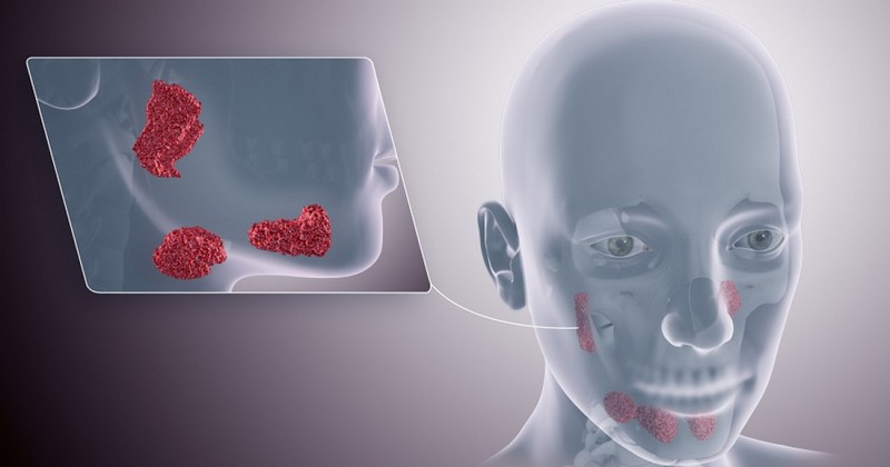
Salivary glands: types, characteristics and functions.

An overview of the salivary glands, important structures in various functions of the body.
On average, an adult human being produces 1 to 2 liters of saliva every 24 hours. The most obvious functions of this liquid are the lubrication of food and the oral cavity, but it has many more hidden properties, all of which are essential for the survival of the living beings that synthesize it.
For example, saliva contains various compounds with Antibacterial properties to prevent colonization of the oral mucosa by unwanted pathogens. Lysozyme is a cationic protein that damages the cell walls of bacteria, rendering the microorganisms inactive. Lactoferrin and peroxidase enzyme also have similar properties, as they cause the destruction of the harmful microorganism by different mechanisms.
Beyond its clear bactericidal action, saliva also acts as a pH buffer (helps protect oral tissues from overly acidic or alkaline foods), participates in the formation of the acquired film and promotes remineralization of dental structures, among other things. As you can see, the functionality of this fluid encompasses much more than simple softening of the food bolus.
Based on these premises, we see special interest in investigating the structures that allow us to continuously synthesize saliva. Today we tell you all about the salivary glands and their function and their functionality in different areas of human physiology.
What are the salivary glands?
First of all, it is necessary to emphasize that salivary glands are tissues of an exocrine nature, i.e., they discharge directly into an internal cavity, the lumen of an organ or the surface of an organism, in this case the mouth.in this case the mouth.
They differ from endocrine glands in that they do not discharge their substances into the bloodstream, as is the case of endocrine glands with hormones and other compounds of a plasma nature.
Classification of their different types and functions
Humans have three pairs of major salivary glands (one on each side of the face) and about 600-1000 minor glands. The most important at a histological level are those included in these three pairs, which respond to the following designation: submandibular gland (SMG), sublingual gland (SLG) and parotid gland (PG). Here are the particularities of each of these tissue formations.
1. Submandibular gland (SMG)
The pair of submandibular glands is located under the floor of the mouth.. Each weighs about 15 grams (about the size of a walnut) and contributes about 60% of the unstimulated salivary secretion, that is, that portion of the fluid that we synthesize without realizing it and without responding to a specific environmental element, such as a pleasant smell or the sight of an appetizing food.
Each of the two submandibular glands is divided into a superficial and a deep lobe, clearly differentiated by the intersection of the mylohyoid muscle. The salivary secretions are drained to the floor of the mouth by Warthon's duct, which clearly flows into the sublingual caruncles, located on each side of the frenulum.
2. Sublingual gland (SLG)
The two sublingual glands are the smallest of the essential secretory trio, weighing only about 2 grams.The submandibulars have 15 submandibulars, compared to 15 submandibulars. In addition, their structure is much more diffuse and they are not encapsulated, as is the case in the other pairs.
Depending on their size and weight is their salivary production rate. They are responsible for only 3-5% of the total saliva secretion, a very small value. They are located just below the sublingual mucosa (hence their name), on the floor of the mouth, resting directly on the anterior lingual surface of the mandible, more specifically on the mililary muscle.more specifically on the mylohyoid muscle.
These glands drain saliva through 8 to 20 excretory ducts, called ducts of Rivinus. The largest of these is the major sublingual (Bartholin's) duct, which is responsible for secreting much of the saliva produced by these diffuse (but essential) small glands.
3. Parotid gland (PG)
Undoubtedly, the two parotid glands are the queens of salivary secretion and the most important within this whole terminological conglomerate. These are the largest glands, weighing between 14 and 28 grams on average and measuring up to 3.4 centimeters in the ventrodorsal axis. They are the producers of approximately 50% of the total saliva produced by the human being, i.e., 0.5 to 0.5 grams.They are the producers of approximately 50% of the total saliva produced by humans, i.e. 0.5 to 1 liter per 24 hours, which is an average of 0.5 to 1 liter per 24 hours.
Up to 80% of the glandular body of each of these structures rests on the external part of the masseter muscle, whose main function is to elevate the jaw and allow the proper closing of the mouth. The remaining 20% of this structure extends medially through the stylomandibular tunnel, which contains a ligament (stylomandibular ligament) that separates the parotid gland from the submandibular gland, among other things.
This glandular tissue is serous in nature, i.e., it focuses on the secretion of polypeptide-like compounds. The cells that make up serous glands (such as this one) possess characteristic zymogen granules, inactive enzyme precursors. The nuclei of these serous cells are rounded, and their cytoplasm has a large amount of rough endoplasmic reticulum (RER).
We are not going to focus too much on the histological section of this gland, as it is too complex. It is enough for us to know a few brief brushstrokes, such as the following: the parotid gland has four faces, lateral, superior, anteromedial and posteromedial.. In addition, it has three defined borders, anterior, medial and posterior. Different structures pass or interact through the parotid, among which are the facial nerve, the retromandibular vein, the external carotid, the superficial temporal artery and the maxillary artery, among others.
It should be noted that this is the area most prone to the appearance of salivary tumors, due to its size and functionality. Without going any further, 85% of salivary gland tumors are located in one of the parotid glands, while the sublingual glands account for only 1% of neoplasms.. In any case, up to 80% of these tumor growths are benign and of adequate development and treatment, so they do not pose a danger to the patient's life.
4. Minor salivary glands
As mentioned above, there are three easily distinguishable salivary gland pairs in the human oral anatomy: the submandibular, sublingual and parotid glands. In any case, there are some 600-1,000 glandular structures also related to the oral cavity, which do not fall into any of the three morphological groups previously mentioned.. These are the minor salivary glands.
These small glandular structures are located under the mucosa of the oral cavity, palate, paranasal sinuses, pharynx, larynx, trachea and bronchi. However, are much more numerous in the buccal, labial, palatal region and in the lingual tissue.. They are also known as accessory glands and are composed of small clusters of saliva-producing acini.
As a whole, they produce less than 10% of the saliva secreted by a human being in a 24-hour interval. Nevertheless, their function is essential, as their continuous production allows oral tissue to remain lubricated and healthy. They also promote tissue safety and prevent bacterial infestation, through the production and secretion of bactericidal salivary compounds that we have already described.
Summary
As you can see, covering the entirety of the salivary glands is very simple, as you only need to have a clear central idea: there are three pairs of major salivary glands (submandibular, sublingual and parotid) and about 1,000 small acini scattered throughout the oral tissue, which are responsible for secreting saliva continuously and uninterruptedly throughout the life of the individual.
Saliva is not only essential for softening the food bolus during and after mechanical chewing, but it also prevents infections, aids in tooth repair and maintenance, acts as a buffer for acid and alkaline pHs, and many other things. and much more. Undoubtedly, it is clear that each and every fluid secreted by the human being has a specific and irreplaceable function.
Bibliographical references:
- Amano, O., Mizobe, K., Bando, Y., & Sakiyama, K. (2012). Anatomy and histology of rodent and human major salivary glands-Overview of the japan salivary gland society-sponsored workshop-. Acta histochemica et cytochemica, 45(5), 241-250.
- Carubbi, F., Alunno, A., Cipriani, P., Bartoloni, E., Baldini, C., Quartuccio, L., ... & Giacomelli, R. (2015). A retrospective, multicenter study evaluating the prognostic value of minor salivary gland histology in a large cohort of patients with primary Sjögren's syndrome. Lupus, 24(3), 315-320.
- Hernando, M., Martín-Fragueiro, L., Eisenberg, G., Echarri, R., García-Peces, V., Urbasos, M., & Plaz, G. (2009). Surgical management of salivary gland tumours. Acta Otorrinolaringologica (English Edition), 60(5), 340-345.
- Martinez-Madrigal, F., & Micheau, C. (1989). Histology of the major salivary glands. The American journal of surgical pathology, 13(10), 879-899.
- Tucker, A. S. (2007, April). Salivary gland development. In Seminars in cell & developmental biology (Vol. 18, No. 2, pp. 237-244). Academic Press.
(Updated at Apr 14 / 2024)