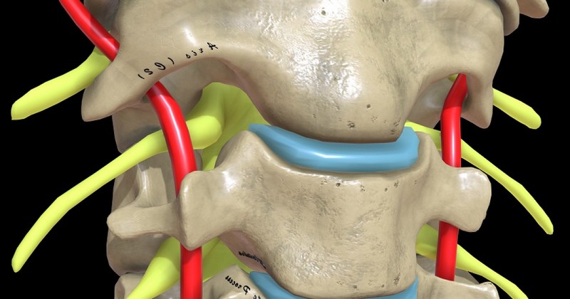
Spinal nerves: what they are, types and functions in the body.

A summary of the characteristics of the spinal nerves, and how they function.
The spinal cord is a long and fragile tubular structure that begins at the end of the brainstem and continues until it almost reaches the final segment of the spinal column. Its main function is to transmit signals and commands created in the brain to the trunk, neck and 4 extremities (efferent function) and in turn to collect all sensations and perceptions recorded throughout the body and send them to the brain (afferent function).
Understanding life without the spinal cord is really complex, and proof of this are the patients with lesions in some part of this fragile but essential structure. Depending on the site of the trauma, from the legs to the whole body may experience a total (complete) or partial (incomplete) loss of sensation and motor capacity below the neurological level of the injury.
Undoubtedly, we could define the spinal cord as the transmission center of all information in the body. It is a neurological highway, whose task is to emit and receive signals to each and every part of our organism with a specific physiological objective. To accomplish this task, the spinal cord is not alone: It has 31 pairs of spinal nerves whose function is to innervate the entire body plane (except the head).. Here we tell you the most relevant information about them.
What are the spinal nerves?
As we have hinted in previous lines, the spinal nerves or spinal nerves are those that extend from the spinal cord and cross the vertebral muscles to distribute to all areas of the body. are those that extend from the spinal cord and pass through the vertebral muscles to be distributed to all areas of the body..
The skeletal muscles of our body are innervated by both motor and sensory nerves, whose function is to collect and transmit the information to the central nervous system (CNS), from where an effector response is generated. This Muscle group comprises more than 600 muscles that can move at will, and together they comprise the muscular system. The smooth and cardiac musculature is outside this motor conglomerate, as the movements they perform are not conscious and are produced "automatically".
Thus, the spinal nerves are directly related to this muscular portion, so that the movements and the development of the human being in a three-dimensional environment are possible.. It should be noted that each of these nerves emerges through the spaces of the vertebrae in the form of 2 short branches, called spinal nerve roots. Here is a brief description of their particularities.
1. Motor nerve root
This root, located in the anterior part of the spinal cord, is responsible for transmitting impulses from the spinal cord to the skeletal muscles to promote their contraction and thus the production of movement..
Radiculopathies (lesions or damage to one or more nerves and their roots) usually cause a characteristic weakening of the muscles innervated by the affected motor root. They become weak, atrophic, flaccid and fasciculated.
Sensory nerve root
On the other hand, the sensory root enters the posterior part of the spinal cord. The nerve fibers that compose it carry sensory information, which will ultimately be interpreted by the brain.. Examples of this information are the position of the body, the degree of luminosity, touch, environmental temperature and pain when suffering an injury, among many other exogenous and endogenous parameters.
As a result, damage to the sensory nerve roots results in a lack of sensitivity in the areas innervated by the injured nerves. Due to this "double" composition of the spinal nerves, it is said that they fulfill a function of a mixed nature: they send and collect information equally.
The 31 pairs of spinal nerves
Yes, you read that right. 31 pairs of spinal nerves emerge from the spinal cord and innervate practically the entire body, except for the head and certain sections of the neck.. The cephalic work is relegated to the cranial nerves, which are 12 nerve pairs whose function is to connect the brain with the eyes, ears, nose, throat and various parts of the head and neck.
Below, we present you the functionality of all the spinal nerves by blocks, as these are divided based on the structures they innervate. Let's go to it.
1. Cervical nerves (C1-C8)
These are the nerves of the first 7 cervical vertebrae. They arise from the spinal cord, emerge through the foramina of the spinal column and are distributed through specific sensory and motor areas..
The cervical nerves innervate the sternohyoid, sternothyroid and omohyoid muscles. In general, these muscle groups can be defined as fleshy ribbons extending from the sternum/ shoulder blade to certain parts of the neck. As a curious fact, it should be noted that the first cervical nerves lack posterior roots in 50% of people.
2. Thoracic nerves (T1-T12)
There are a total of 12 spinal nerves that emerge from the thoracic vertebrae.. Almost all of them are located between the ribs (intercostal), with the twelfth located below the last rib (subcostal nerve). The intercostal nerve endings are distributed throughout the walls of the thorax and abdomen.
These thoracic nerves participate in the functions of the organs and glands of the head, neck, thorax and abdomen. They are responsible for the innervation of the mammary glands, chest wall, abdominal wall and pelvis. Due to their importance at the nervous level, these spinal nerves are the therapeutic targets of choice for many treatments aimed at managing chronic pain in patients.
3. Lumbar Nerves (L1-L5)
These are 5 spinal nerves arising from the lumbar vertebrae. They are divided into 2 compartmentalized sections, anterior and posterior. These nerve elements emerge from the spine through the foramina of conjunction.. However, these nerves should not be conceived as a series of isolated entities: the first 3 and most of the fourth are connected to each other in this situation by anastomotic loops, forming the lumbar plexus.
Thus, the lumbar plexus is established between the anterior branches of the L1 and L4 spinal nerves. On the other hand, the smaller part of the fourth nerve joins with the fifth nerve to form the lumbosacral trunk, which participates in the formation of the sacral plexus.
4. Sacral nerves (S1-S5)
These are the 5 spinal nerves that emerge from the sacral bone (bone located below the lumbar vertebra L5 and above the coccyx) and constitute the lowest segment of the spinal cord.. Although the vertebral components of the sacrum are fused to form a single bony entity, each of these nerves is named after the vertebra to which it would be associated.
These nerves are divided into branches, but many of them end up joining each other, and also the lumbar and coccygeal plexuses. As previously mentioned, this series of interconnections form plexuses, specifically the sacral and lumbosacral. Branches of these plexuses innervate the hip, thigh, leg and foot.
5. Coccygeal nerve
The coccygeal nerve is the last of the spinal nerves, i.e. number 31. It arises in the conus medullarishelps form the coccygeal plexus and innervates the sacrococcygeal joint and part of the levator ani.
8 cervical nerves + 12 thoracic nerves + 5 lumbar nerves + 5 sacral nerves + 1 coccygeal nerve: 31 spinal nerves.
Summary
In this space, we have covered the general particularities of the 31 spinal nerves that run throughout our body, except for the head and certain parts of the neck. Their function is to emit information from the brain and allow muscle contraction (motor work) and, in turn, to receive all the essential information provided by the extremities and innervated areas (sensory work).
Thanks to these spinal and cephalic nerve pairs, human beings are able to function in a three-dimensional environment, being aware of our own internal state and of what surrounds us in the environment. After reading these lines, one concept is clear to us: without our nerve endings, human beings are nothing.
Bibliographical references:
- Alzola, M. S. R. (2002). Nervous Tissue.
- Diseases of the spinal cord, Medlineplus.gov. Retrieved March 5 from https://medlineplus.gov/spanish/spinalcorddiseases.html.
- Maza-Marrugo, M. P. (2020). Nerve fibers, peripheral nerves, endings. Neuroanatomia, 10.
- Nerves, Merck Manual. Retrieved March 5 from https://www.merckmanuals.com/es-us/hogar/enfermedades-cerebrales,-medulares-y-nerviosas/biolog%C3%ADa-del-sistema-nervioso/nervios.
- Spinal nerves or spinal nerves, Dolopedia. Retrieved March 5 from https://dolopedia.com/categoria/nervios-espinales-o-nervios-raquideos.
- Spinal nerves, Fisio Online. Retrieved March 5 from https://www.fisioterapia-online.com/glosario/nervios-raquideos
- Rodríguez-García, P. L., Rodríguez-Pupo, L., & Rodríguez-García, D. (2004). Clinical techniques for the neurological physical examination. I. General organization, cranial nerves and peripheral spinal nerves. Rev Neurol, 39(8), 757-66.
- Romero, L. V. (2015). Anatomy and physiology of the nervous system. XinXii.
- Willard, F. H., & KEY, C. (2006). Autonomic nervous system. Ward RC, editor. Fundamentals of Osteopathic Medicine. 2nd ed. Buenos Aires: Editorial Médica Panamericana, 94-125.
(Updated at Apr 12 / 2024)