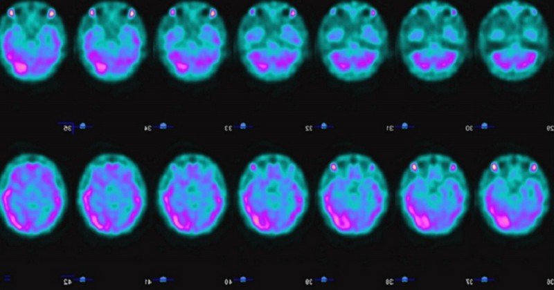
Brain Spect: what is it and how does this neuroimaging method work?

The technological solution to many cases in which it is necessary to know the state of the brain.
Neurological evaluations are diverse. There is a wide range of methods that can be used to determine whether our brain is functioning properly or whether there is any abnormality.
The technique called spect cerebral is a method which allows us to see images of the functioning of specific parts of the brain by means of substances that are injected into the subject's organism.
In this article we will review the main characteristics of this evaluation technique, we will see in which cases it is applicable and its contribution in the pharmacological field.
What is brain spect? Characteristics
The spect brain is a neurological evaluation tool that consists mainly of injecting a substance intravenously, which adheres to specific brain structures depending on their chemical composition, and allows the evaluator to observe how that particular area is functioning.
This is possible because the substance injected into the body has a detection mechanism by means of radioactive isotopes, which are added to the body.which are added to the patient's body intravenously. Prior to this, a gamma radiation source must be applied to the subject. Once this substance is in the patient's body, it is mixed with his Blood until it reaches the brain, and it is there where it adheres to the structure that the specialist needs to evaluate. As mentioned before, the chemical composition of the substance will determine to which specific structure of the brain it adheres.
This method, also known as single-photon emission computed tomography, is extremely practical to performIt does not require any complex preparation. It is only the application of gamma radiation to the patient to later perform the intravenous injection into the organism. Then the substance is responsible for making the tour and show the areas of interest.
The estimated duration of this method is approximately one hour, calculating the whole process of asepsis prior to the application.
What does it evaluate?
Basically, there are three aspects that this test allows to evaluate. It is the study of cerebral perfusion, tumor viability and cerebral receptors.
Cerebral perfusion
It is evaluated by means of radioisotopes, which depending on the level of the patient's blood flow, will be fixed in the encephalic tissue.. This procedure provides significant information on vascular pathologies that are difficult to detect with other tests.
In addition, it is also effective in indirectly showing the activity of neurons. This aspect is of great importance in the field of psychiatry.
Tumor viability
This is done using tracers that do not perforate the blood vessel network, which remains intact. These tracers are actively incorporated into the subject's organism as potassium analogs.
The importance of this evaluation lies in determining tumor conditions or natural changes in the organism as a result of surgery..
3. Neuro-receptors
Finally, this analysis makes it possible to evaluate the density and distribution of the different receptors in the Central Nervous System (CNS).. It is achieved thanks to emitting isotopes specially marked for the procedure.
This aspect is the most recent in terms of spect cerebral evaluations. In spite of this, it has shown a fairly good degree of efficiency when required.
In which cases is it applied?
This form of evaluation has been shown to be extremely useful in a wide variety of cases; it is even able to detect neurological and psychiatric abnormalities that other techniques miss.
Some of its most frequent uses are in cases where the extent of cerebrovascular disease (CVD), Parkinson's disease, dementia in all its forms, and epilepsy need to be assessed. In these assessments, brain spect is extremely effective. It is also able to recognize areas of the brain that have a lower than normal blood supply.This is a very effective form of prevention for cerebrovascular disease.
With regard to epilepsy, this evaluation technique can capture by means of the photogram the irritative focus during the seizure, which helps to know exactly which brain area is affected and to proceed with the necessary intervention.
With regard to psychiatric illnesses, it is of great help in establishing the differential diagnosis between in establishing the differential diagnosis between disordersIt also provides information in the recognition of multiple brain areas affected and the need for intervention. It also provides information in the recognition of multiple more complex neuropsychiatric pathologies.
Contributions to pharmacology
In the field of pharmacology, the brain spect has been very useful, helping to determine which drugs are more efficient at the time of its iteration with the nervous system, especially of neurotransmitter inhibitor drugs..
Taking into account that this technique allows to clearly see how is the path of the drug in the organism, the level of blockage towards a certain substance and when its effect can last before a new dose is necessary.
Bibliographic references:
- Dougall NJ, Bruggink S, Ebmeier KP (2004). "Systematic review of the diagnostic accuracy of 99mTc-HMPAO-SPECT in dementia". Am J Geriatr Psychiatry. 12 (6): 554 - 570.
- Scuffham J. W. (2012). et al. "A CdTe detector for hyperspectral SPECT imaging. Journal of Instrumentation. IOP Journal of Instrumentation. 7: P08027
(Updated at Apr 12 / 2024)