What are tendon calcifications?
The calcifications are a disorder characterized by calcium crystal deposition that harden a fabric with elastic characteristics such as tendons, muscles, fasciae, or bursae. In this article we will focus specifically on the tendon pathology and its calcification as one of the most abundant reasons for. Tendinopathies affect a large part of the population between 40 and 60 years of age and its symptoms are pain in the area of the injury, muscle weakness or even bone deformities under the skin if there is calcification.
Possible causes of tendon calcification
Calcifications appear by a ongoing inflammation in the same area caused by repetitive movements or microtrauma. It is a injury associated with aging of the body and work activity. In fact, tendinopathies are defined as occupational injuries because they are caused by strained postures and repetitive movements at work. The tissue is repeatedly injured and fails to regenerate again, so it is replaced by low-resistance hard tissues or hydroxyapatite crystal deposits that end up forming a small bone in the tendon.
Some of the factors predisposing to suffer this type of injury are:
- Microtrauma and mechanical overload.
- Aging of tissues with decreased blood flow.
- Inadequate nutrition.
- Problems derived from a malfunction of the organism.
- Genetic causes.
Where are the most common ones located?
As previously discussed, calcifications are the result of repeated inflammation. They are that have become chronic and are located in joint structures such as the shoulder, elbow, ankle, etc. The most frequent injuries are:
- Rotator cuff: are the muscles that stabilize the shoulder and its tendons attach to the humerus. Due to the great mobility of the joint, the arrangement of the tendons and the activity that it can endure on a daily basis, it is an area that tends to continuously become inflamed, thus predisposing the appearance of calcifications in its tendons. The most common lesion is calcification of the supraspinatus. Rotator cuff tendinopathies cause difficulties in lifting the arm or performing rotations such as combing or fastening a bra.
- Calcaneal spur: It's about a bone growth on the bottom of the heel of the foot, exactly in the calcaneus bone where the musculature of the sole of the foot and the fascia have insertion. Sometimes in the radiological images a thickening of the calcaneus bone can be seen in the part of the sole of the foot in a pointed way. The pain is located in the back part of the sole of the foot and makes it difficult to support while walking.
- Achilles tendon: it is the tendon of insertion of the twins and the soleus in the most posterior and superior part of the calcaneus bone. Calcifications appear due to muscle overload or repetitive tendinitis. The pain is located in the area of insertion of the Achilles tendon and can expand to the calf and the sole of the foot.
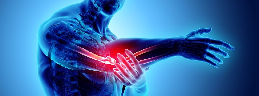
- Humerus epicondyle: the tendons of the forearm muscles are attached to the elbow, exactly at the two epicondyles, internal and external. The tendons tend to become very inflamed due to the load suffered by the forearm muscles, causing the famous '' '' ''. Its complication involves the appearance of calcifications in the area, making it impossible to actively flex or extend the arm.
Diagnosis of calcifications
It is diagnosed through a X-ray or ultrasound. pain, inflammation and functional impotence are the main symptoms. They are usually diagnosed as tendonitis initially and treated as such. When the imaging tests and the radiological signs of calcium deposits in the soft tissue are evident, it is when it is diagnosed as a calcification and an evaluation of a new treatment is carried out to eliminate them if necessary. Sometimes calcifications can stay dormant and discovered when performing an imaging test for a different reason, which indicates that you can also have a calcification and not have any symptoms.
Treatment according to the type of tendon calcification
Its treatment during acute episodes is the application of local ice, anti-inflammatories, rest and physical therapy. There are treatments conservatives (electrotherapy devices and manual therapy) or invasive like surgery. If the calcification has been diagnosed by a radiological image and the pain persists for a long time, more specific treatments are proposed to remove the calcium deposits from the area. Next, we will detail what are these treatments.
Conservative treatments:
Invasive treatments:
yasmina Santiago
-
Electrotherapy: several can be used electrotherapy devices in the case of calcifications that will help the destruction and reabsorption of calcified tissue and the regeneration of healthy tissue. It is usually combined with manual therapy.
- Shock waves: they are waves that are applied with a conductive head on the skin in the area where the calcification is located and fragment the calcium deposits, thus promoting their reabsorption by the same organism.
- Iontophoresis: it involves the application of local analgesic medication through currents applied to the skin to reduce pain and inflammation.
- Low power laser: it is applied with a head on the skin that emits a low-power laser in order to activate tissue regeneration by stimulating cellular response.
- Corticosteroid infiltrations: If the pain persists and is very intense, this more invasive treatment can be used in the area of the injury.
-
Arthroscopy: Applies when conservative treatment has not worked and pain has persisted for more than six months. A camera, instruments, and an aspirator are inserted into the joint through small incisions to break up and clean the calcified tissue. It is a simple operation, but it also requires a postsurgical treatment based on a physiotherapy rehabilitation program to reduce pain, accelerate tissue recovery and joint functionality.
- Calcifications are the result of repeated inflammation. They are tendinitis that have become chronic and are located in joint structures such as the shoulder, elbow, ankle, etc.
- Its treatment during acute episodes is the application of local ice, anti-inflammatories, rest and physical therapy.
- If the pain persists and is very intense, there are more invasive treatments such as corticosteroid injections in the region of the injury.
(Updated at Apr 14 / 2024)