What is choriocarcinoma and how is it treated?


Choriocarcinoma is form of cancer, in most cases associated with pregnancy. It develops from germ cells (cells of the embryo), which form its outer shell and ensure the immersion of the embryo into the uterine mucosa and the formation of the placenta (child's place). These cells are capable of unlimited division and growth into maternal tissues. Their growth is restrained by special mechanisms in the mother's organism during gestation: the immune system, the decidual membrane (loose mucous membrane of the uterus involved in the formation of the placenta) of the uterus, etc.
In case of violation of protective mechanisms, as well as when elements of the embryo enter the blood and beyond the mucous membrane of the uterus, the development of choriocarcinoma is possible.
This type of cancer is distinguished by high malignancy and is quickly metastasizing (spread to other organs), more often in the vagina and lungs.
Rare forms of chorionic carcinoma are not associated with pregnancy and develop in girls and women who have not previously become pregnant from ovarian germ cells (teratoblastoma).
Short information about gestational choriocarcinoma
- Stage 1 - cancer within the uterus;
- Stage 2 – cancer dissimilates beyond the uterus, but only to the genitals;
- Stage 3 cancer - metastasizes to the lungs;
- Stage 4 - cancer extends to distant organs (other than the lungs).
Choriocarcinoma symptoms

- Serous (transparent) discharge from the genital tract, eventually becoming purulent.
- Bloody discharge from the genital tract of varying intensity (from smearing to profuse). They do not respond to hormonal treatment and do not go away after curettage of the uterus.
- Bleeding from other organs with metastatic lesions (spread of tumor cells to other organs and growth of daughter tumors in them) of the lungs, brain, etc.
- Pain of varying intensity in the lower belly or organs affected by daughter tumors (metastases).
- Deterioration of general well-being;
- Increased body temperature;
- Significant decrease in body weight;
- Loss of appetite;
- Nausea;
- Severe weakness, fatigue and decreased performance.
Incubation period
In most cases, choriocarcinoma develops within three to twelve months after giving birth, miscarriage, or abortion, but the development of the disease is possible even 10-20 years after pregnancy, including in postmenopausal women (after age-related cessation of menstruation (monthly uterine bleeding associated with physiological endometrial rejection - inner layer of the uterine mucosa)).
Forms

According to the degree of spread of the tumor process, choriocarcinoma is divided into stages:
- Stage 1 - cancer within the uterus;
- Stage 2 – cancer dissimilates beyond the uterus, but only to the genitals;
- Stage 3 cancer - metastasizes to the lungs;
- Stage 4 - cancer extends to distant organs (besides the lungs).
Causes

The reasons are not fully understood. It is believed that the development of chorionic carcinoma is facilitated by:
- Viral infection during pregnancy;
- Decreased immunity, in which tumor cells are not recognized or destroyed by the body, which contributes to their engraftment and growth in various organs and tissues;
- Ingress of elements of embryonic tissues (for example, after an abortion) with blood flow outside the uterine lining, for example, into the vagina or fallopian tubes, where the germ cells begin to divide intensively and turn into malignant.
Risk factors:
- Complicated course of the preceding pregnancy (spontaneous abortion, artificial abortion, ectopic pregnancy (development of pregnancy outside the uterine cavity), etc.);
- Cystic drift (a disease that develops during pregnancy, characterized by abnormal growth of the embryonic part of the placenta in the form of bubbles that penetrate deeply into the wall of the uterus);
- Early onset of sexual activity (up to 15 years);
- Late onset of menstruation (monthly uterine bleeding associated with physiological rejection of the endometrium - the inner layer of the uterine lining): the first menstruation after 15 years.
Diagnostics

- Analysis of the anamnesis of the disease and complaints (when (how long ago) there were pains in the lower abdomen, discharge from the genital tract).
- Analysis of obstetric and gynecological history (past pregnancies, their outcomes, abortions, gallbladder drift (a disease that develops during pregnancy, characterized by abnormal growth of the embryonic part of the placenta in the form of bubbles that penetrate deeply into the wall of the uterus) ) etc.).
- Analysis of menstrual function (at what age the first menstruation began (monthly uterine bleeding associated with physiological rejection of the endometrium - the inner layer of the uterine mucosa), what is the duration and regularity of the cycle, the date of the last menstruation, etc.).
- Gynecological examination with obligatory bimanual (two-handed) vaginal examination. The gynecologist with both hands by touch (palpation) determines the size of the uterus, ovaries, cervix, their ratio, the state of the ligamentous apparatus of the uterus and the area of the appendages, their mobility;
- Ultrasound examination (ultrasound) of the abdominal cavity and small pelvis through the anterior abdominal wall and ultrasound with a transvaginal transducer. With the help of these examinations, it is possible to determine the location and size of the tumor, the involvement of the surrounding tissues in the process. An addition to the ultrasound examination is color Doppler mapping (CDM). The method allows you to determine the nature of blood flow or its absence in the tumor, which makes it possible to confirm the diagnosis of malignant formation.
- Magnetic resonance imaging (MRI) of the brain, abdominal cavity is performed to exclude metastases (daughter tumors) to these organs.
- Computed tomography (CT) is performed to exclude metastases to the brain, lungs, and abdominal organs.
- Hysteroscopy (a special device - a hysteroscope - is inserted into the uterine cavity through the vagina and the cervical canal): the study confirms the presence of a tumor in the uterine cavity. During hysteroscopy, a section of the tumor (biopsy) is taken for examination under a microscope - a histological examination.
- Diagnostic curettage of the uterine cavity, followed by histological examination of the resulting tissues to search for tumor cells.
- Consultation with an oncologist is also possible.
Choriocarcinoma treatment

- Polychemotherapy of a tumor - treatment of choriocarcinoma with a group of drugs (cytostatics, for instance Hydroxyurea) that can inhibit the growth and destroy tumor cells.
- Surgical treatment - removal of the tumor when:
- Large size of the uterus (corresponding to 9-10 weeks of pregnancy);
- The danger of rupture of the uterus or ovary by a rapidly growing tumor;
- Massive bleeding.
- Surgical treatment (removal of the uterus, ovaries, and fallopian tubes) is also used for primary choriocarcinoma (teratoblastoma) in girls and non-pregnant women, if the tumor does not respond (does not decrease in size, the level of chorionic gonadotropin (a hormone synthesized by the tumor) does not decrease) to chemotherapy.
Complications and consequences
- Compression of neighboring organs when reaching large sizes (up to 10-15 cm in diameter): the appearance of false desires to empty the intestines, frequent urination, pulling pain in the abdomen.
- Uterine bleeding.
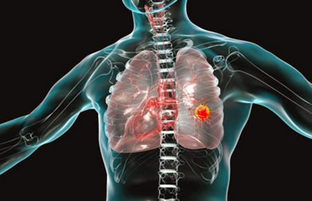
- Metastases to the lungs, which is manifested by hemoptysis (coughing up blood), chest pains, coughing, shortness of breath (feeling short of breath, rapid breathing).
- Vaginal metastases, accompanied by bloody and purulent discharge from the genital tract.
- Metastases to the liver develop when choriocarcinoma is large, accompanied by pain in the right hypochondrium. Can lead to rupture of the liver and severe bleeding into the abdominal cavity.
- Brain metastases are characterized by intense headaches, nausea, vomiting that does not bring relief, loss of sensation or immobilization (paralysis) of parts of the body.
Despite the pronounced aggressiveness, with timely diagnosis, the tumor is treatable. With early and adequate treatment, the prognosis for life and fertility is favorable. The advanced forms with metastases are severe and can be fatal.
Choriocarcinoma prevention
- Rational nutrition with a sufficient content of selenium, vitamin A, a low amount of animal fats.
- Quitting bad habits (smoking, alcohol abuse).
- Planning for pregnancy (exclusion of abortion).
- Regular visits to the gynecologist (2 times a year).
Post by: Katherine Christensen, gynecologist, sexologist, Copenhagen, Denmark
(Updated at Apr 14 / 2024)
Hydrea articles:
Some of the trademarks used in this Web Site appear for identification purposes only.
All orders are reviewed by a licensed physician and pharmacist before being dispensed and shipped.
The statements contained herein are not intended to diagnose, treat, cure or prevent disease. The statements are for informational purposes only and is it not meant to replace the services or recommendations of a physician or qualified health care practitioner. If you have questions about the drugs you are taking, check with your doctor, nurse, or pharmacist.