What is chronic compression myelopathy and how is it treated?


Compression myelopathy is a very serious complication of the nervous system diseases. It is a compression of the spinal cord by various formations: disc herniation, hematoma, tumor, the formation of bone fragments in any trauma.
The main symptom of the disease is the loss of sensory and motor function. In addition, the work of internal organs is disrupted. To diagnose this disease, computed tomography, as well as myelography and radiography are performed. Spinal cord compression must be treated promptly.
Brief information about the disease
This term means damage to the spinal cord due to pressure on it due to something with a violation of the sensory and motor systems. It is not an independent disease. This is a complication that occurs in pathological processes in the spinal membranes or in the spinal column.
This complication is caused by the violation of the substance by pathological factors, such as clamping of large blood vessels, because of this, the nutrition of the nervous tissue is disturbed, and also, necrosis develops. The longer this complication lasts, the more intensively the blood flow changes.
Main types and reasons of the disease development
Depending on the rate of development of spinal cord compression, myelopathy is divided into the following types:
- Acute. The form of the disease develops with simultaneous compression of the brain substance, which at the same time damages its structure. And it is just as pronounced as neurological symptoms. This occurs from the moment of injury to clinical symptoms. The cause of this condition is hemorrhage of the lining of the spinal cord or spinal injury. It can also represent an epidural abscess or the outcome of a tumor process.

There are compression fractures of the vertebrae - this is the same reason that can become an acute syndrome of compression myelopathy, as a result of which fragments are displaced. This occurs due to the large load on the spine, for example, when diving in an unfamiliar place, hitting the bottom with your head hard.
In absolutely all of the aforementioned cases, the spinal cord is clamped in the spinal canal or compressed by bone fragments. Spinal cord hemorrhage can occur due to taking medications that reduce blood clotting activity in any back injury. The spinal cord is located in the bony canal, which is formed from holes in the vertebral body, and is surrounded by membranes on all sides. Blood from a vein of a damaged vessel is poured between the hard spinal membrane and the bone. The blood is not able to compress, and the spinal canal is narrow enough, a hematoma forms there, which pushes the spinal cord, as a result of which it squeezes.
- Subacute (occurs over 1-2 weeks). Such compression myelopathy occurs when a purulent process forms, rapid tumor growth and rupture of an intervertebral hernia. Such tumors and complications may not appear for a long time, and not make themselves felt. But when the nervous tissue no longer compensates for all the damage, subacute compression of the spinal tissues will eventually develop. The same happens during the formation of suppuration under the membrane. When purulent edema begins to grow in size, then compression and damage to the spinal cord occurs.
- Chronic. The main feature is the gradual growth of compression myelopathy, for example, over many years.
Symptoms of compression myelopathy
The manifestations of the disease directly depend on the type of compression and the location of the injury site. The spinal cord is not a homogeneous structure. At the very beginning, motor neurons are located in the spinal structure, they are responsible for muscle movement, and at the end are nerve cells. On the sides there are centers responsible for the work of internal organs.

The symptoms that appear depend on where the compression occurs. All three types of myelopathy form differ in the rate of development, symptoms of the disease, and functions that will soon be lost.
Acute compression is the most difficult to tolerate. With it, the motor system falls out, that is, flaccid paralysis develops, and the sensitive system of functions of a part of the body also "falls out". If the lower section is damaged, then the work of the rectum and bladder is disrupted. This condition is called "spinal shock".
After some time, this flaccid paralysis turns into spastic, that is, convulsive contractions of various muscles, pathological reflexes appear, and, possibly, the development of contractures (joint stiffness).
Compression in the cervical (neck) spine
Compression myelopathy begins with dull pain in the neck, upper chest, back of the head, as well as shoulders and arms. In these areas, disorders of the sensory system begin to appear, this manifests itself in the form of a feeling when goosebumps creep, numbness of some parts of the body. After that, the stage of muscle fatigue begins, and a decrease in their tone, as well as atrophy. Twitching of some muscle fibers is sometimes observed.
If compression occurs in the first and second cervical segments, then the damage of the facial nerve (violation of sensitivity on the face) is inevitable. In addition, hand tremors and unsteady gait are possible.
Compression in the chest spine
At this site compression occurs very rarely. This includes weakness in the legs, as well as decreased sensitivity in the back, abdomen, and chest.
Compression myelopathy in the lumbar (lower back) spine
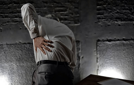
Such compression is characterized by pain in the thighs, muscles of the buttocks, lower leg. This also degrades the sensitivity of these areas. The more time passes, the more the muscles begin to lose tone, weakness appears, and atrophy also occurs. In one or both legs, flaccid peripheral paresis eventually occurs.
Diagnostics of myelopathy
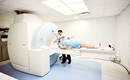
The most common diagnosis is magnetic resonance imaging or computed tomography of the spine. On pictures of the spinal cord, you can see the sites of damage, the condition of the spinal cord, and even the reasons of the violations.

If there is no opportunity to undergo tomographic diagnostics and especially if there is a suspicion of dislocation of the vertebrae or a fracture, radiography in three projections is usually used. A lumbar puncture is also performed, which examines the cerebrospinal fluid. A special method can be used, it is based on the introduction of a coloring contrast into the subarachnoid space - this is myelography. When this substance has dispersed, a picture is taken. And thanks to them, a doctor can determine at what level the spinal compression had happened.
There are varying degrees of myelopathy. Below is the European Myelopathic Scale. See how you are doing:
European Myelopathic Scale
Classification by score
Treatment of myelopathy
Acute and subacute myelopathy requires immediate treatment. The purpose of such treatment is, as soon as possible, to remove the spinal element from which compression and damage occurs. It helps reduce damage to nerve pathways. Chronic compression requires immediate intervention. Treatment depends on the period when the compression occurred, as well as its magnitude.

Usually, for a chronic disease caused by osteochondrosis, a two-stage treatment regimen is proposed. Initially, a course of therapy is prescribed. This stage includes taking vitamins, anti-inflammatory drugs (first of all, NSAIDs such as Nimesulide and others), drugs to reduce muscle spasm (muscle relaxers) such as Tizanidine, and other drugs that help repair tissue.
If these methods do not help, then you cannot do without surgical treatment. The following methods can be carried out:
- Removal of a vertebral hernia;
- Laminectomy (surgical removal of the back of one or more vertebrae to give access to the spinal cord or to relieve pressure on nerves);
- Facetectomy (surgery remove the pressure of the spinal nerve roots);
- Disk replacement with a prosthesis;
- Removal of the tumor, cyst, or bone growth (in spondylosis) causing compression;
- If there is stenosis of the spinal canal, then microsurgical dilation of the spinal canal is done;
- In some cases, an additional plate or vertebral body replacement is needed.
Keep in mind that even after surgery additional physiotherapeutic procedures and certain changes in a lifestyle are required.
The most important role in treatment is played by continuous therapy, that is, annual rehabilitation courses, in special medical institutions. Doctors prescribe to patients daily healing gymnastics that must be chosen individually for each patient by an exercise therapy doctor.
Prevention of myelopathy
Acute myelopathy is the most severe of the three forms. It is severe in all its clinical manifestations. If treatment is started on time, the chances of recovery increase. The reason for the favorable recovery is that the muscles and peripheral nerves do not have time to undergo profound changes. Therefore, with such treatment, it is possible to quickly recover and return functions that have been lost.
In chronic myelopathy, irreversible changes occur in the spinal cord, that is, tissues grow, muscle atrophy occurs. That is why even if the patient immediately begins his treatment, it will be impossible to replenish the sensory and motor functions. According to statistics, serious complications in spinal cord compression occur due to improper treatment and diagnosis.
Conclusion
It's always important to listen to your body. So be your own inner doctor. So, if you have been experiencing severe back pain in certain areas for a long time, or even functional failures or false sensations, then consult a doctor immediately. This should be a spine specialist, neurosurgeon, or podiatrist.
When it comes to surgery, you have to think about the good, namely, about 90% of all patients improve. Improvement is, of course, most evident when the problem is detected earlier and dealt with in a timely manner. Since today modern spinal surgery operations are performed more sparingly and quickly, the patient is mobilized the next day after the operation.
It is also comforting that the disease no longer occurs in the operated part and ensures the improvement of the condition of the affected segment of the spine.
However, prevention is better than any treatment. Here are a couple of tips:

- Avoid one-sided loading;
- Avoid long-term improper posture.
- Move regularly.
- Strengthen your back and abs.
- Even for mild complaints, seek the help of a medical-gymnastics specialist.
- Avoid prolonged flexion work and do not pinch the phone between your ear and shoulder.
- If you are working in front of a monitor, try to sit so you could look straight whenever possible.
Post by: Emma Ager, MD, Copenhagen, Denmark
(Updated at Apr 15 / 2024)
Zanaflex articles:
Some of the trademarks used in this Web Site appear for identification purposes only.
All orders are reviewed by a licensed physician and pharmacist before being dispensed and shipped.
The statements contained herein are not intended to diagnose, treat, cure or prevent disease. The statements are for informational purposes only and is it not meant to replace the services or recommendations of a physician or qualified health care practitioner. If you have questions about the drugs you are taking, check with your doctor, nurse, or pharmacist.