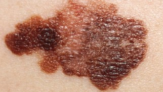
What is melanoma – symptoms, treatment, prognosis


Melanoma is a cancerous tumor that emerges from the pigment cells of melanocytes. This cancerous tumor most often starts on the skin or, most commonly, a birthmark. Rarely, localization occurs in other organs, such as the eyeball, oral mucosa, or the subungual space. It is one of the most dangerous human malignant tumors, which is characterized by frequent relapses and metastatic lymphogenous and hematogenous processes.
Short information about melanoma
Clinical forms of malignant melanoma:

- Surface spreading. In women, it usually occurs on the legs, and in men – on the back and chest. Superficial formations develop at a slow pace, gradually spreading over the skin.
- Nodal. Affects the upper body.
- Acral Lentiginous. Affects palms or under nail skin.
- Lentiginous (Hutchinson's malignant freckle). It develops against the background of a pigmented spot (birthmark). According to statistics, elderly women are more susceptible to this form. Localization - the skin open to the sun, commonly, it is a face. It is characterized by horizontal, slow growth and has the most favorable outlook.
- Achromatic (non-pigmented). The rarest type of melanoma.
Melanoma can affect the eyes, nose, mouth, lungs, rectum, etc. Therefore, there is a classification of internal disease occurence:
- Retinal melanoma;
- Cancer of the mucosa;
- Malignant melanoma of soft tissues.
Also, there is the concept of progressive melanoma. In this case, cancer cells travel from their original site to other organs, causing another tumor called secondary or metastatic cancer. It can affect the digestive tract, central nervous and respiratory system.
Sometimes malignant melanoma occurs even many years after the elimination of the primary formation.
Etiology (causes)

The main provocative factors for the disease is uncontrolled exposure to solar radiation or artificial ultraviolet radiation (tanning bed). Excessive enthusiasm for beach or tanning bad entails a violation of the DNA pattern of skin cells and increases the likelihood of malignant process.
There are also additional risk factors, among them:
- Phenotype. Individuals with a light skin tone, blondes, as well as those with blue eyes, are more prone to this disease;
- Weakened immunity;
- Large number of moles;
- A history of sunburn;
- Pigmented xeroderma (genetic disorder making skin unable to fully repair UV damage);
- Age group over 50;
- Heredity. If more than two family members have a similar disease, you should pay special attention to your moles and undergo examinations regularly.
- The presence of nevi (dark moles) (a melaniform nevus is especially dangerous);
- Previously diagnosed melanoma.
Pathogenesis
The stage of the disease is based on the depth and dissimilation of cancer cells. The penetration depth is determined by the methods of Breslow and Clark.
Melanoma stages (according to Breslow):

- I - the formation occurs only in the superficial layers of the skin (in the epidermis, up to the basement membrane; thickness less than 2 mm).
- II – cancerous cells are found in the superficial layers of the skin (deeper than 2 mm; may be characterized with an ulcer).
- III - the cancer process disseminates to the adjacent lymph nodes and/or goes beyond the primary tumor site.
- IV, V - cancer cells are located in the deeper layer of the skin, grow into adipose tissue, metastases spread throughout the body, with selective damage to organs.
Subdivision of Clark into microstages includes thin, intermediate, and thick (deep) invasions. Projections vary based on existing stages of melanoma.
If the disease is early diagnosed and treated timely (stages I, II), then the prognosis is excellent and is 98.2% survival rate. Stage III leaves the chances of life within 61.7%. IV, V stages (especially with metastasis) are characterized by a rapidly progressive and fatal course. As a result, less than 10% survive for five years.
Clinical manifestations
Most types of malignant melanoma begin with skin changes or a new mole. Also, old mole, which has changed its shape, is in danger.
Signs of melanoma to note when testing moles:
- Geometry - an asymmetrical mole, in which one half is not equal to the other;
- Border - a mole whose contour is not rounded, clarity disappears;
- The color is uneven and consists of several shades;
- Diameter exceeds 5 mm.
Itching or bleeding are additional symptoms of skin cancer. They are less common but must not be ignored.
Also, you should pay attention to:
- Hair loss from the surface of the nevus;
- Ulceration;
- A sharp increase in size;
- Nodulation.
Complications after primary removal
Symptoms of progressive melanoma may appear many years after the initial mass is removed. Manifestations are based on the resources of the human body and organs affected by cancerous cells. A secondary formation can appear in the form of tumors on the skin, lungs, or any organs of an individual. Additional signs include a sharp decrease in body weight, loss of appetite, fatigue and exhaustion.
Melanoma diagnostics

During the diagnosis, it is necessary to carefully examine moles for signs of the disease. It is crucial to examine all areas of the skin - the scalp, body folds, between the toes, feet, and the lumbar region. An independent visual inspection is advised to be repeated at least once every six months, it is even better to contact a specialist who will conduct a professional inspection.
Instrumental methods for diagnosing melanoma include:
- Dermatoscopy -exam of the skin using a specialized apparatus. As a result, the degree of danger of the disease is determined, with the possibility of assessing the development of events. Dermatoscopy is an essential step in detecting skin problems.
- Any suspicion of skin cancer requires a biopsy. The taken tissues from the affected area with further histology should confirm or deny the presence of an oncological problem.
To identify the stage of the process, the oncologist prescribes additional tests, such as laboratory blood tests, ultrasound, computed tomography, etc.
Treatment

The first stage is characterized by a melanoma depth smaller than 1 mm and no signs that raise the risk of relapse, such as an open wound or multiple mitosis. Removal of melanoma covers an area with a diameter of 1 cm.
Trherapy of stage I and II melanoma. If cancerous cells are found deeper than 1 mm, a biopsy of the lymph nodes is performed. If lymph nodes are intact, local surgery will solve the problem.
If adjacent lymph nodes are affected, then stage III has begun, which requires a more extensive operation. In the therapy of stage III melanoma, patients are offered an auxiliary immunological therapy.
Actions in stages IV, V, when cancer dissimilates through the body and forms metastases, require a combination of surgical intervention and other methods. The therapy is contingent on the site of the cancerous growth, general health, and previous treatments for the tumor. Biochemical and immunological therapy are the most common methods for solving the problem. Radiation therapy can help control symptoms in case of spread to the central nervous system, digestive tract, or respiratory system.
As for the surgical intervention, it is made to eliminate the tumor and adjacent lymph nodes. After a biopsy confirming the diagnosis, extensive local resection is performed. The surgeon cuts off adjacent tissues to make sure no cancerous cells are left behind. It also lowers the risk of melanoma relapse.
There are also additional medicines used at home, for instance, Hydroxyurea, a drug that disrupts the growth, development and division mechanisms of all cells in the organism, including malignant ones, thereby initiating natural cells death.
Healing control
After removal of the tumor and depending on the degree of the disease, follow-up is planned. Patients with early-stage melanoma should see a doctor every month. Any changes in the area of operation require immediate professional advice. During routine check-ups, doctor checks the adjacent lymph nodes. As part of self-control, it is necessary to pay attention to changes in moles, if they appear.
After surgery and deep therapy of melanoma, constant consultation with an oncologist is required, who will monitor the condition. During follow-up, additional tests such as x-rays and blood tests may be appointed.
Prevention of melanoma

After any treatment for malignant melanoma, it is crucial to avoid uncontrolled sun exposure and to take precautions to protect the skin, such as:
- Wearing cotton or natural fabrics that provide better sun protection;
- Always wearing a wide-brimmed hat in the sun;
- Wearing sunglasses and choosing a shade for relaxation and movement;
- Use of sunscreen with high reflectivity (spf);
- Being in the sun at a safe time (before 10:00 and after 16:00).
Tips and tricks for coping with the disease psychologically

1. Adapt to the diagnosis, talk to a psychotherapist.
2. Cope with the change in lifestyle.
3. Analyze the causes of the disease.
4. Constantly maintain hope for a cure.
5. Be sure to inform your family.
6. Continue work if possible.
7. Taking short, regular walks improves your well-being.
The patient must accept and adapt to changes in life. Treatment can change your daily routine and cause physical discomfort, side effects (hair loss, nausea, vomiting, weight loss, fatigue and weakness). In most cases, these effects go away after therapy end. The consequences of melanoma are not as terrible as it seems to the patient - in the event that you seek qualified medical help in time.
What doctor treats melanoma?
If your family physician finds signs of melanoma, they will refer you to a narrow profile specialist. Further treatment is carried out directly by the dermatologist (in case of confirmed diagnosis - oncologist). In many cases, a dermatologist can distinguish a benign growth from a cancerous tumor during a preliminary examination of a mole. In this case, the complex will be assigned with the necessary diagnostic procedures to determine the degree of damage and the appointment of appropriate treatment. It will not be superfluous to get an opinion from another specialist to confirm the diagnosis and determine the methods of control.
Post by: John Johansson, M.D., Amsterdam, Netherlands
(Updated at Apr 14 / 2024)
Hydrea articles:
Some of the trademarks used in this Web Site appear for identification purposes only.
All orders are reviewed by a licensed physician and pharmacist before being dispensed and shipped.
The statements contained herein are not intended to diagnose, treat, cure or prevent disease. The statements are for informational purposes only and is it not meant to replace the services or recommendations of a physician or qualified health care practitioner. If you have questions about the drugs you are taking, check with your doctor, nurse, or pharmacist.