What is traumatic brain injury (craniocerebral trauma)?


Craniocerebral trauma or traumatic brain injury (TBI) is a medical term for contact injuries caused by external effect that affect soft tissues of the head, skull and brain, having a single mechanism and duration of formation, i.e. caused at the same time.
TBI is can be closed and open. With an open TBI, the skin is damaged, the aponeurosis and the bottom of the wound is bone or deeper tissues. Moreover, if the dura mater is damaged, then an open wound is considered to be penetrating. In a closed TBI, the aponeurosis is not affected but the skin is damaged.
Short information about traumatic brain injury (craniocerebral trauma)
Clinical forms of TBI
- Skull fracture - Fractures are more often bone-linear.
- A concussion is the most frequent type of TBI. It accounts up to 75% of all registered TBIs. It is associated with mild impairment of neurological function. All symptoms that occur after a concussion usually disappear over time (within a few days - 7-10 days). If they persist for more time, it means that there is a more serious brain damage. A concussion may or may not be followed by fainting. Nonspecific symptoms include nausea, vomiting, pallor of the skin, cardiac abnormalities. Neurologic examination usually shows no abnormalities, but somatic symptoms (headache), physical signs (fainting, memory loss), behavioral changes, cognitive impairment, or sleep disorder may occur. Some of these effects can last for several months after the trauma.

- Brain contusion. It can be mild, moderate and severe (clinically). It is a more severe form of TBI. The same symptoms are accompanied by the pronounced focal violations such as paresis (paralysis of certain groups of muscles), speech disorder and others as a result of softening of the medulla. Brain contusion manifests itself in a bruised wound of the brain. The bruised wound occurs in the place of the hit and then the bruised wound is inflicted on the contrary side of the brain. Clinical manifestations depend on the site of the injury, and include a change in mental state, differently dilated pupuls, increased drowsiness, confusion, agitation. Small intraparenchymal hemorrhages and edema of the adjacent tissue can often be identified with computed tomography.
- Impact and counter-impact injury.
- Diffuse axonal injury. It is severe injury to the axonal white matter of the brain as a result of shear force caused by strong acceleration or inhibition of the brain.
- Compression of the brain.
- Intracranial hemorrhage.
There could be various combinations of types of TBI in one patient: contusion and compression by hematoma diffuse axonal damage and contusion, and so on.
Symptoms

- External hematomas, tissue ruptures;
- Pallor;
- Vomiting, nausea;
- Irritability;
- Lethargy, drowsiness;
- Weakness;
- Paresthesia;
- Headache;
- Impaired consciousness - loss of consciousness, somnolence, stupor, coma, amnesia, confusion;
- Neurological signs - convulsions, ataxia;
- The main indicators of the state of the body may change - deep or arrhythmic breathing, hypertension, bradycardia - which may indicate increased intracranial pressure.
TBI can be primary (due to injury) and secondary (due to internal complications) by localization - focal and diffuse; by the nature of damage and depth - closed, open and penetrating, non-penetrating; and by severity mild, moderate, or severe.
Instrumental examination
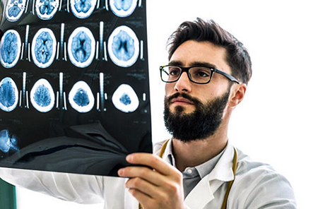
For penetrating and blunt traumatic brain injuries, non-contrast CT is more commonly used. It has sufficient sensitivity to detect acute hemmorhage and skull fractures. CT also allows assessing the severity of the trauma in detection of elevated intracranial pressure, brain swelling and dislocation. The following CT data may indicate the threatening nature of the injury: displacement of the midline structure, smoothing of the sulcus, enlarged or compressed ventricles, loss of normal differentiation of gray and white matter.
Laboratory research
The main tests for TBI are general blood test, an analysis of electrolytes is performed. Glucose control should be performed in children with impaired consciousness. Patients with intracranial bleeding should have a coagulogram (prothrombin time, activated partial thromboplastin time).
Treatment
Examination and treatment for serious injury should be done at the same time. Treatment of TBI can be divided into 2 stages. The stage of first aid and the stage of providing qualified medical care in the hospital.

The initial examination is carried out with special attention to the patency of the airways, breathing, immobilization of the neck part of the spine.
In fainting, even a single episode, the patient, regardless of their current state, needs to be transported to the hospital. This is due to the high potential risk of developing severe life-threatening complications.
After admission to the hospital, the patient undergoes a clinical examination, a history is collected, if possible, and the nature of the trauma is clarified with them or with those accompanying. Then a set of diagnostic measures is performed aimed at checking the integrity of the bone skeleton of the skull and the identification of internal hematomas and other effect on the brain.
After the type of TBI is identified during, the neurosurgeon or the traumatologist decides on the need for surgery and control of intracranial tension. The aim of the first stage of medical help is to avoid secondary brain damage, for which oxygenation and gas exchange are maximized, blood circulation is maintained to maximize cerebral perfusion, elevated intracranial tension and the metabolic needs of the brain are lowered.
Severe TBI often requires intubation to protect the airway.
In some cases, trepanation and drainage of intracranial hematomas are made to avoid damage to brain, to maintain normal pressure and protect the cerebral cortex from hypoxia (insufficient oxygenation). If there is no bleeding into the cranial cavity, patients are usually treated with conservative therapy. Premedication can be done with etomidate, a sedative that is neutral to the cardiovascular system and has minimal effect on blood pressure. Most other sedatives result in hypotension, and hence a lowering in arterial tension and a drop in cerebral tension.
Rehabilitation after TBI
The treatment itself depends on the severity of the injury and can consist of both taking medications and surgical intervention. During rehabilitation, the patient is monitored by a doctor and using X-ray, CT, MRI, ultrasound and other examinations.
Rehabilitation takes place individually: some patients need to restore motor skills, and some need to get rid of headaches, improve sleep, etc. The doctor selects a set of procedures based on the tasks that are in front of him.
Rehabilitation is divided into several periods:
1. Early: 2-5 days after injury. The doctor determines the severity of the injury, its possible consequences (headache, ringing in the ears, dizziness, nausea, vomiting) and the presence of complications (fainting, amnesia, sleep disturbance, emotional lability).
2. Intermediate: 5-30 days after injury. The tasks of rehabilitation are to train the vestibular apparatus, restore mobility to the limbs, if necessary, reduce spasticity (movement disorders caused by increased muscle tone. Usually, muscle relaxers such as Tizanidine are used). Rehabilitation measures include: drug treatment, breathing exercises, physiotherapy, massage, thermotherapy (cryo-, paraffin therapy), reflexology.

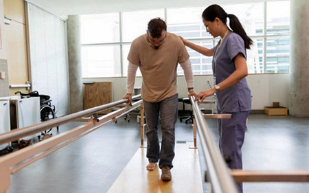
3. Late recovery: 1-4 months after injury. If necessary, the patient continues therapy, but the intensity of the loads, like the medications taken, can be changed by the doctor. Physical therapy is actively used to restore impaired motor functions and reduce hypertonicity. Respiratory gymnastics allows adjusting the ventilation of the lungs. To train balance, the patient works with supports, performs special physical exercises, oculomotor gymnastics. If necessary, the doctor deals with the patient with the restoration of household and work skills.
4. Residual: up to two years after injury. During this period, the main functions are usually restored, but if the patient still experiences some difficulties, the doctor continues to work with the patient, restore motor skills, speech, fine motor skills, coordination of movements and self-care skills using exercise therapy, occupational therapy, training on simulators, etc. The rehabilitation process is impossible without massage: it helps to establish blood and lymph circulation, muscle tone, restore motor functions, and relieve pain. Physiotherapy and thermotherapy fight spasticity.
Consequences of traumatic brain injury
Depending on the severity of the injury, the patient may experience: impairment of memory and concentration, depression, bouts of unreasonable anxiety, decreased performance, physical abilities, rapid fatigue, dizziness, headache, partial or complete loss of vision, sleep problems, impaired breathing, blood circulation, speech, physical activity, epilepsy, etc. All the consequences can bother the patient for many years, even if the injury was not severe.
In severe injuries the possible consequences can be seen almost immediately after hospitalization. Unfortunately, this type of injury is one of the leading causes of deaths, disability, coma, and vegetative state in young people.
Post by: Kylie Richardson, General Practitioner, Rotterdam, Netherlands
(Updated at Apr 14 / 2024)
Zanaflex articles:
Some of the trademarks used in this Web Site appear for identification purposes only.
All orders are reviewed by a licensed physician and pharmacist before being dispensed and shipped.
The statements contained herein are not intended to diagnose, treat, cure or prevent disease. The statements are for informational purposes only and is it not meant to replace the services or recommendations of a physician or qualified health care practitioner. If you have questions about the drugs you are taking, check with your doctor, nurse, or pharmacist.