Angioresonance: Revolutionizing Vascular Diagnostics

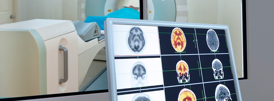
Magnetic resonance angiography (MRA) is the study of blood vessels through the use of magnetic resonance imaging. It is a non-invasive technique, unlike usual angiography, during which a catheter must be inserted into the patient's blood system to be able to observe the structure of the vessels and their possible alterations. Magnetic resonance imaging is an imaging test that is based on subjecting the patient to a magnetic field and radio frequency waves that stimulate the body's protons and, after stimulation, they return a signal that is collected by a computer that transforms it into images in two or three dimensions of the analyzed structures.
Advantages and limitations
The advantages of MRA over arteriography and CT angiography are that it is a non-invasive examination and that it does not subject the patient to ionizing radiation; it is a quick procedure, very little bothersome and cheaper than the other tests. Also, gadolinium practically never causes allergic reactions, unlike the iodinated contrast used in CT angiography.
On the contrary, MRA has its limitations, since it does not allow the visualization of calcium deposits within the arteries and the precision of the images of certain vessels, especially those of smaller caliber, is not as exquisite as when angiography is performed. conventional. In turn, the quality of the images obtained by MRA can be affected and be of low quality if the patient is not able to remain still and lying down for the duration of the test.
In which situations it applies
The MRA application is very useful when assessing the following situations:
- Aneurysms of the aorta or other arteries
- Involvement of the carotid arteries that supply the brain
- Brain arteriovenous malformations
- Peripheral vascular disease in the lower and upper extremities
- Renal Artery Involvement in the Evaluation of a Kidney Transplant
- Evaluation of the arteries supplying a tumor before your surgery
- Coronary Artery Assessment Before Stenting or Bypassing
- Study of congenital arterial malformations
- Preventive study of patients with relatives who have suffered aortic aneurysms
How to prepare the patient
MRA requires the same preparation as a normal MRI. In some cases, a contrast, called gadolinium, may need to be injected to enhance some images and better assess certain structures. In such cases, the patient will be asked to go on a 4-6 fasting period and the contrast will be injected intravenously. The test can take between 20 and 60 minutes. During it, the patient is placed lying down inside the device, which is a closed cylinder that can cause claustrophobia in certain people. Sometimes it can be useful to give a pain reliever before the test and during the test there is always a button at hand that can be pressed to stop the study if the patient needs to stop for a few minutes. Loud noises will be heard while ARM is being carried out and may be annoying, but acoustic protection is always provided. It should be said that nowadays there are open MRI machines, reducing the feeling of claustrophobia that can be experienced during the test and that they are also useful in the case of very large people.
Do you have any contraindications?
MRA subjects the patient to a magnetic field, so this test is contraindicated if the patient has a cardiac pacemaker or defibrillator, cochlear implants, surgical clips used in brain aneurysms, or metal coils placed inside vessels blood vessels to expand them. Most metals used in prosthetic joints, artificial heart valves or nerve stimulators do not contraindicate the use of an MRA, but the procedure should be avoided during the recent postoperative period, for at least 6 weeks. Dental fillings are not affected by the magnetic field, but they can distort the images of the cranial region.
MRA is not recommended for a pregnant woman during the first trimester of pregnancy, unless strictly necessary. However, it has not been documented that subjecting a pregnant woman to MRA can cause harm to the fetus. If she is breastfeeding and contrast has to be applied, the patient is recommended to express her milk a few days before and not to breastfeed again until 48 hours after the test to have eliminated the contrast.
- Non-invasive technique, unlike the usual angiography that uses magnetic resonance to study the blood vessels.
- It is fast, very little annoying and inexpensive, although it has some limitations, especially in terms of image quality.
- Aneurysms of the aorta or other arteries, cerebral arteriovenous malformations, peripheral vascular pathology in the arms and legs are some of its most useful applications.
REMEMBER THAT: In addition to the most beneficial guarantees included in each modality, they offer insured persons additional benefits and advantages that protect them against any mishap related to their well-being and that of their family.
(Updated at Apr 13 / 2024)