Bronchoscopy

Alternative names
Fiberoptic bronchoscopy.
Definition of bronchoscopy
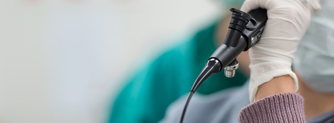
Bronchoscopy is a diagnostic and / or treatment test that consists of visualizing the airways (larynx, trachea and larger bronchi) through the use of a bronchoscope or rigid or flexible hollow tube that has an adapted optical system and that is connected to a monitor or a computer in which the images obtained are recorded.
How is the study done?
The study is carried out in adapted units or operating rooms by the doctor or specialized personnel in aseptic conditions and generally on an outpatient basis.
During the study the patient remains seated (sometimes lying) on an armchair in a comfortable way, with the head directed forward and the chin slightly inclined downwards. The patient is asked to breathe normally through the mouth while the examiner slowly inserts the flexible bronchoscope (shaped like a hollow tube) through one nostril (sometimes the bronchoscope is inserted directly through the mouth); You are then asked to begin to breathe through your nose until the bronchoscope is directed through the pharynx, larynx, trachea, and main bronchi at the same time that the explorer visualizes the airways. Tweezers can be inserted through the orifice of the bronchoscope to collect tissue samples or an aspiration system can be connected to obtain samples of secretions. During the bronchoscopy study, sedatives or local anesthetics are usually used to inhibit the nausea or vomiting reflex and minimize the discomfort of the procedure. At the same time vital signs and oxygen saturation are recorded during the study.
The use of the rigid bronchoscope requires the use of general anesthesia, it is always placed through the mouth and is reserved for very specific examinations or for taking biopsies.
The study usually lasts 15-20 minutes.
Preparation for the study
The patient must avoiding smoking and taking exciting substances such as caffeine 48-72 hours prior to the study. You should avoid the intake of solid and liquid food for at least 6-8 hours before the scan.
Before starting the examination, they are removed.
Some medications must be withdrawn days prior to the study, especially anticoagulants, anti-inflammatories or antiaggregants.
What does it feel like during and after the study?
The study is annoying but not painful for the patient thanks to the use of anesthetics and sedatives.
During the study, a sensation of nausea, coughing or slight pressure may occur during the placement of the bronchoscope.
The patient should avoid the intake of liquids or solids until the effect of the local anesthesia has worn off (between one or two after the study is carried out) to avoid the passage of food or liquids to the respiratory tract.
Hours or days after the bronchoscopy is performed, the patient may present mild nasal, oral or respiratory discomfort such as itching, burning, or mild pain that can be relieved with commonly used analgesics or anti-inflammatories. Coughing spells may appear accompanied by mucus with small traces of blood.
The patient should consult with his doctor in the case of fever, nose or mouth bleeding, difficulty breathing, swallowing or speaking, in the hours or days after the study.
Study risks
- allergy to drugs used during the study
- Lesions of the oral or respiratory mucosa
- Nose or mouth bleeding
- Vocal cord injury
- Breathing difficulty (rare)
- Viscus Piercing (Rare)
- Risks inherent to the use of general anesthesia (in studies that require its use)
Study contraindications
The patient should consult with his doctor before carrying out the study in the case of:
- allergy to the drugs used in the study
- Taking medication
- Lung disease, heart, blood clotting or high blood pressure, among others.
Reasons why the study is carried out
Bronchoscopy is a widely known and used diagnostic and / or treatment test in the field of Medicine.
At a diagnostic level, it allows the study of patients with chronic cough, respiratory distress, emission of bronchial secretions, active bleeding from the mouth, etc., and identify their possible causes. On the other hand, it allows to directly visualize lesions that have been previously identified in imaging studies (such as X-ray, ultrasound or resonance, among others) and to take tissue or secretion samples for later study.
At the therapeutic level, it allows the extraction of foreign bodies from the airway that cause obstruction to the airway, dilate narrowed bronchi, aspirate secretions or administer different treatments directly on the airway.
(Updated at Apr 14 / 2024)