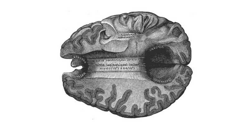
Corpus callosum of the brain: structure and functions

This part of the brain acts as the main bridge linking the two cerebral hemispheres.
Let us think for a moment about a human brain. It is a highly complex structure in which there are two clearly differentiated parts, the two cerebral hemispheres.
We also know that each of these hemispheres has some more specialized functions in different aspects, such as speech, for example.For example, speech is generally in the left hemisphere, or we have seen that while the right hemisphere is more holistic or global, the left hemisphere is more logical and analytical. However, these two hemispheres are not loose and separated from each other, but at some point in the anatomy of the brain it is possible to find a point of union. This junction point is the so-called corpus callosum..
What is the corpus callosum?
The main bundle of nerve fibers that connects both cerebral hemispheres is called the corpus callosum. This structure is mainly made up of neuronal axons coated with myelin, thus forming part of the white matter of the brain. myelin-coated neuronal axons, thus forming part of the white matter of the brain. Within the white matter, the corpus callosum is considered an interhemispheric commissure, since it connects and exchanges information between structures of the different hemispheres. There are other interhemispheric commissures in the human brain, but they are much smaller than the corpus callosum.
This structure is located in the midline of the brain, lying at the bottom of the interhemispheric fissure, and being mostly hidden from external observation by being partially covered by the cortex. It is leaf-shaped or comma-shaped, possessing different parts that connect differentiated parts of the brain to each other.
The areas connected by this structure of the brain are mostly cortical areas, with some exceptions. Usually subcortical structures connected to other structures and commissures.
Parts of the corpus callosum
Although the corpus callosum is considered a single structure, it has traditionally been divided into several parts. Specifically, the corpus callosum could be divided into the following four sections.
1. Peak or rostrum
Located in the inferior frontal part of the corpus callosum, this is the most anterior part of this structure. It arises from the lamina terminalis and is connected to the optic chiasm.
Genu or knee
This is the part of the corpus callosum that curves towards the interior of the brain, heading anteriorly towards the frontal lobes to form the minor forceps. The fibers of this part of the corpus callosum connect the prefrontal cortexes of the two hemispheres, allowing their information to be integrated..
3. Body
Posterior to the genu or knee, is the body, which ends thickening in its posterior part. It connects with the septum and the trigone.This in turn is an important connecting structure between regions of the brain, such as the thalamus, the hippocampus and other areas of the limbic system.
4. Splenium or buckle
The most posterior and final part of the corpus callosum is formed by the fibers that end up associating with other projection and associative fibers. It connects with the occipital lobe to form the greater forceps, and also connects to the lateral ventricle. is linked to the lateral ventricle to the point of forming one of its inferior walls.. It also connects to the pineal gland and the habenular commissure (which connects the habenular nuclei of both hemispheres).
Functions of this part of the brain
The main function of the corpus callosum is to transmit information from one hemisphere to the other, enabling interhemispheric communication.allowing interhemispheric communication. Thus, the fact that the functions of each of the hemispheres are partly different does not prevent them from acting as an integrated whole, allowing the precise execution of the different processes and actions carried out by the human being.
In this sense, it is also is also linked to learning and information processing, by linking and acting as a liaison between the hemispheres.It is also linked to learning and information processing, by uniting and acting as a link between the different brain nuclei. On the other hand, if for example a part of a cerebral hemisphere is injured, thanks to the corpus callosum the opposite hemisphere can take care of those functions that are left unattended.
In addition, some studies show that apart from this function, the corpus callosum also influences vision. also influences vision, specifically eye movement.This is natural, as the information about the eye muscles is transmitted through it. It is natural, since in eye movements the coordination between the two hemispheres, in this case the eyes, is crucial.
What happens when it is sectioned?
The corpus callosum is an important structure for integrating the information received and processed by both cerebral hemispheres. Although the absence of a connection between hemispheres at the level of the corpus callosum does not imply a complete loss of functionality (since the corpus callosum is the main inter-cortical commissure between the hemispheres). Although it is the main interhemispheric commissure, it is not the only one.), the total or partial disconnection of the cerebral hemispheres can be a major handicap for the performance of various activities.
Among other things, this kind of disconnection between parts of the brain can give rise to what is known as callosal disconnection syndrome.
In this syndrome, patients with split brain (i.e., with a disconnection between the two hemispheres) have shown difficulties such as incoordination, repetition or perseveration in performing sequenced activities. difficulties such as incoordination, repetition or perseveration in performing sequential activities such as combing, feeding or dressing such as combing, feeding or dressing, sometimes performing the same action twice due to lack of motor integration.
Also learning and retention of new information is also greatly hindered by the inability to coordinate by not being able to coordinate information correctly (although it does not make it impossible, it requires a much greater effort than usual), as well as it can cause alexia (inability to read) and agraphia (inability to write).
In addition, significant alterations can occur at the sensory level. For example, it has been shown that posterior lesions of the corpus callosum can cause severe difficulties in discriminating between somatic stimuli, causing somatic agnosias or lack of recognition from tactile stimuli.causing somatic agnosias or lack of recognition from tactile stimuli. Memory and language problems are also common.
Callosotomy: when sectioning the corpus callosum can be a good thing.
Despite the disadvantages that this kind of surgical intervention may entail, in the presence of some very serious disorders, the division of the corpus callosum or callosotomy has been evaluated and successfully applied for medical purposes as a lesser evil. for medical purposes, as a lesser evil.
The most typical example is that of resistant epilepsy, in which the sectioning of parts of the corpus callosum is used as a method of reducing severe epileptic seizures, preventing epileptoid impulses from traveling from one hemisphere to another. In spite of the problems it may cause by itself, callosotomy increases the quality of life of these patients, since the difficulties it may cause are less than those produced by continuous comict crises, thus reducing the risk of death and the quality of life may even improve.
On the other hand, with time it is possible that the brain reorganizes itself to allow mental processes that during the first weeks after the operation seemed to be eliminated or seriously damaged, although recovery is not usually complete.
Conditions affecting the corpus callosum
It has been previously indicated that the division of the corpus callosum may have limiting effects, although sometimes its section may be considered in order to improve the symptomatology of a disorder.
However, However, the corpus callosum being cut or damaged may occur accidentally or naturally.There are multiple diseases that can affect this area of the brain. Some of these alterations may occur as a result of the following.
1. Cranioencephalic traumas
After a blow or trauma, the corpus callosum can be easily damaged mainly due to its great consistency and density. Generally The substance of the corpus callosum is usually torn, or there is diffuse axonal damage.The corpus callosum can be easily damaged, mainly due to its high consistency and density. If we speak of focalized effects in one point, the greatest affectation is usually in the splenium.
2. Cerebrovascular accidents
Although it is not frequent due to the bilateral irrigation that the corpus callosum has, it is possible to find cases in which hemorrhages cases in which hemorrhages or ischemia produce an involvement of the white matter of the corpus callosum.. In this way, alterations in Blood flow are capable of practically cutting off the communication between the two hemispheres that takes place in the corpus callosum, without the need for a solid element to come into contact with this part of the brain and break it.
3. Demyelinating disorders
Being a structure formed by white matter, covered with myelin, disorders such as multiple sclerosis greatly affect the corpus callosum.. This type of disorders causes that the messages sent by the brain are not sent in an efficient way or even that many neurons die, so that in the corpus callosum the perceptions and functionalities of both hemispheres cannot be easily integrated. In this way, mental processes involving regions on both sides of the brain are greatly affected, or cannot be carried out at all.
4. Brain Tumors
Although its compactness means that in general there are not many tumors affecting the corpus callosum, some very aggressive tumors such as lymphoma or glioblastoma multiforme some of great aggressiveness such as lymphoma or glioblastoma multiformewhich is usually located in the white matter, can infiltrate and affect this specific structure and cause serious damage or "strangle" it by the pressure exerted by the growth of the cancerous parts.
In the case of glioblastoma it usually produces a typical butterfly-shaped pattern with greater involvement of the central area.
5. Malformations
Although not very frequent, it is possible to find malformations in some subjects that cause them to have fewer connections than usual from birth. Other types of congenital malformations can cause easy rupture (and consequent hemorrhage). (and consequent hemorrhage) of blood vessels in the brain, which can also affect the corpus callosum.
Bibliographic references:
- Bishop, K.M.; Wahlsten, D. (1997). Sex Differences in the Human Corpus Callosum: Myth or Reality?. Neuroscience & Biobehavioral Reviews. 21 (5): 581 - 601.
- Hofer, S.; Frahm, J. (2006). Topography of the human corpus callosum revisited—Comprehensive fiber tractography using diffusion tensor magnetic resonance imaging. NeuroImage. 32 (3): 989 - 994.
- Kandel, E.R.; Schwartz, J.H. & Jessell, T.M. (2001). Principios de neurociencia. Cuarta edición. McGraw-Hill Interamericana. Madrid.
- Mantilla, D.L.; Nariño, D.; Acevedo, J.C.; Berbeo, M.E. y Zorro, O.F. (2011) Callosotomía en el tratamiento de epilepsia resistente. Universidad Médica de Bogotá, 52(4): 431-439.
- Peña-Casanova, J. (2007). Neurología de la conducta y Neuropsicología. Editorial médica Panamericana.
- Witelson, S. (1985). The brain connection: The corpus callosum is larger in left-handers. Science. 229 (4714): 665–8.
(Updated at Apr 13 / 2024)