Dermoscopy

Alternative names
Epiluminescence microscopy.
Definition
Exploration in dermatology it is eminently visual. To visualize some dermatological lesions, it is of special interest to have tools that facilitate this task and that allow the visualization of skin structures not identifiable to the naked eye.
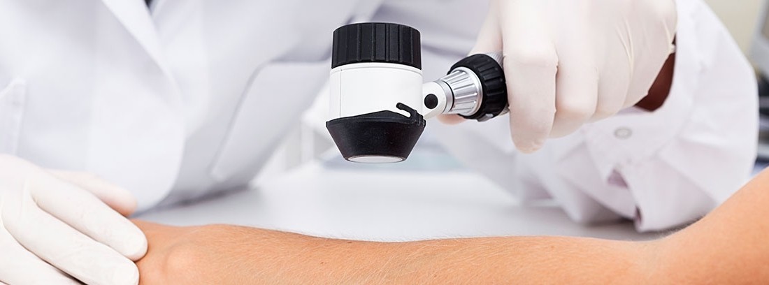
Dermoscopy, although initially developed to improve the diagnosis of melanoma, It is also effective in the evaluation of other skin processes. It is a simple, non-invasive technique that improves the clinical diagnosis of skin lesions, especially pigmented ones, and has become an essential diagnostic technique in the consultation of the dermatologist. The instrument used in this technique is called dermatoscope. Provides an enlarged and sharper image of the dermatological lesion to be studied. It uses a magnification system with a light source that illuminates the skin and allows a magnification of 10 to 400 times. Thanks to dermoscopy, the characteristics of the lesion can be determined and a treatment indicated or the need for some other more complex complementary examination such as biopsy.
When the dermoscopy device is coupled to a computer system, it enables digital monitoring of pigmented lesions, the so-called digitized epiluminescence microscopy. This system allows accurate periodic monitoring of pigmented lesions in patients at high risk of developing melanoma. Record digital photographs to assess the evolution in subsequent controls.
How is the study done?
Dermoscopy is a simple exam performed by the dermatologist in his office. The patient must expose the lesion to be studied to the dermatologist. You will apply the dermatoscope on the lesion and visualize its characteristics. A drop of oil may be applied to the lesion to avoid light scattering, which can sometimes make visual examination difficult. Some devices have a special light, polarized light, which avoids the phenomenon of scattering and therefore does not require immersion oil on the skin, being able to directly contact the lens to the skin surface.
Preparation for the study
Although it does not require any special preparation, it is generally recommended to wear comfortable clothing and with the surface to be explored clean and free of creams or ointments, except when medically indicated. Once the exam is done, the patient can return home.
What does it feel like during and after the study?
It is totally painless. It does not involve discomfort of any kind.
Study risks
It is a safe test that does not pose any risk.
Study contraindications
Without contraindications.
Reasons why the study is carried outDermoscopy has proven usefulness in the study of skin tumors, especially pigmented ones. It should always be used for any lesion of these characteristics, since it facilitates the differential diagnosis and improves the diagnostic precision of melanoma.
It is advisable to follow up some patients to guarantee the early diagnosis of melanoma. Patients at high risk for developing melanoma are considered:
- patients with a total number of nevi greater than 50
- presence of atypical nevi
- patients with a family or personal history of melanoma,
- carriers of risk mutations to develop melanoma
- fair skin with a history of sunburn
(Updated at Apr 14 / 2024)