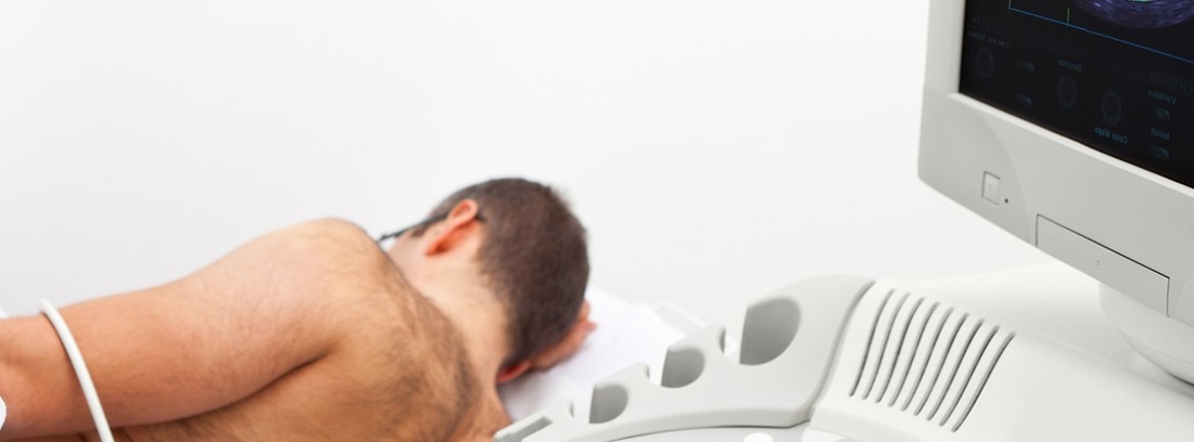
Echocardiogram

Study of the cavities of the heart and its different internal structures through the use of an ultrasound machine which is connected to a monitor or a computer in which the images obtained are recorded for later study.
Depending on how we perform the echocardiography, we can classify it into:
- When the ultrasound is applied directly to the left hemithorax (just above the heart), we speak of transthoracic or conventional echocardiography.
- When the ultrasound is introduced through the mouth and esophagus and reaches the heart area, we speak of transesophageal ultrasound.
The study does not require the use of ionizing radiation (X-rays), contrast or radiopharmaceuticals.
How is the study done?
Echocradiography is performed in the ultrasound room or in the doctor's office at the medical center or hospital.
The patient must bare the chest. It is not necessary to remove jewelry or metal objects as they do not interfere with ultrasound waves. You will lie on a table during the study. The technician will apply a conductive gel on the thorax and will move the ultrasound machine over it to obtain different images of the heart. Once the study is finished, the studied area will be cleaned with a disposable wipe.
In some cases it is necessary to perform a transesophageal echocardiography for a better visualization of the heart, for which the ultrasound in the form of a probe is inserted through the mouth, and it is delivered to the heart area through the esophagus.
The duration of the exam will depend on the findings found during it, but it is usually about 15 minutes.
Preparation for the study
The echocardiographic study does not require prior preparation by the patient. In transesophageal echocardiography, fasting for 6-8 hours prior to the study may be required.
How does it feel to last you and after the study?
Conventional echocardiography is painless. For the patient, local cold may be felt when applying the conductive gel.
Transesophageal echocardiography can be uncomfortable for the patient. A sedative is usually given to prevent nausea and vomiting while the ultrasound is inserted. You may feel slight pressure exerted by the ultrasound scanner during placement.
Study risks
Echocardiography is safe and risk-free because it uses ultrasound waves that are harmless to the patient.
Contraindications to the study
The ultrasound study has no contraindications
What is the study done for?
The echocardiographic study is a simple and safe test, widely known and used in the field of Cardiology since it provides very important information for the doctor about the state of the internal structures of the heart.
It allows the diagnosis and monitoring of alterations such as an increase or decrease in the size of the heart chambers, alterations in the heart valves (which intervene in the correct distribution of blood flow inside them), increase or decrease in the thickness of the walls of the heart (which are involved in the correct distribution of blood throughout the body), and so on.
Transesophageal echocardiography is an invasive test, annoying for the patient, so it is reserved for when conventional echocardiography cannot be performed or does not provide the necessary information.
Echocardiography is a basic test in the field of Cardiology that must be completed with more specific studies in the event of detecting alterations.
(Updated at Apr 13 / 2024)