Glial cells: much more than the glue of neurons

Glia is a substance that outnumbers neurons and is involved in many functions.
It is very common that, when talking about a person's intelligence, we refer specifically to a very specific type of cell: neurons. Thus, it is normal to call mononeurons to those who we attribute low intelligence in a derogatory way. However, the idea that the brain is the idea that the brain is essentially a collection of neurons is increasingly outdated..
The human brain contains more than 80 billion neurons, but this accounts for only 15% of the total number of cells in this set of organs.
The remaining 85% is occupied by another type of microscopic body: the so-called glial cells.. Together, these cells form a substance called glia or neuroglia. form a substance called glia or neurogliawhich extends through all the nooks and crannies of the nervous system.
Currently, glia is one of the most progressive fields of study in neuroscience, in search of unveiling all their tasks and interactions they perform in order for the nervous system to function as it does. The brain cannot be understood today without understanding the involvement of glia.
The discovery of glial cells
The term neuroglia was coined in 1856 by the German pathologist Rudolf Virchow. This is a word that in Greek means "neuronal (neuro) glue (glia)", since at the time of its discovery it was thought that neurons were joined together to form the nerves, and, moreover, that the axon and, moreover, that the axon was a collection of cells rather than a part of the neuron. Therefore, it was assumed that these cells found near the neurons were there to help structure the nerve and facilitate the junction between them, and nothing more. A rather passive and auxiliary role, in short.
In 1887, the famous researcher Santiago Ramón y Cajal came to the conclusion that neurons were independent units and that they were separated from each other by a small space that later became known as the synaptic space. This served to disprove the idea that axons were anything more than parts of independent nerve cells. However, the idea of the passivity of glia remained.. Today, however, it is being discovered that its importance is much greater than previously assumed..
In a way, it is ironic that the name given to neuroglia is that. It is true that it does help in the structure, but it not only performs this function, but they are also for its protection, repair damage, enhance nerve impulse, provide energy, and even control the flow of information, among many more functions discovered. They are a powerful tool for the nervous system.
Types of glial cells
Neuroglia is a collection of different types of cells that have in common that they are found in the nervous system and are not neurons..
There are quite a few different types of glial cells, but I will focus on discussing the four classes that are considered most important, as well as explaining the most prominent functions discovered to date. As I have said, this field of neuroscience is advancing more and more every day and I am sure that in the future there will be new details that are unknown today.
Schwann cells
The name of this glia cell is in honor of its discoverer, Theodore Schwann, best known as one of the fathers of the Schwann Cell Theory.. This type of glial cell is the only one found in the Peripheral Nervous System (PNS), that is, in the nerves that run throughout the body.
While studying the anatomy of nerve fibers in animals, Schwann observed cells that were attached along the axon and gave the impression of being something like small "beads"; beyond this, he gave them no further importance. In further studies, it was discovered that these microscopic bead-like elements were actually myelin sheaths, an important product generated by this type of cell.
Myelin is a lipoprotein that provides insulation against electrical provides insulation against the electrical impulse to the axonMyelin is a lipoprotein that provides insulation against the electrical impulse to the axon, i.e., it allows the action potential to be maintained for longer and at a greater distance, making the electrical impulses go faster and not disperse through the neuron membrane. In other words, they act like the rubber covering a wire.
Schwann cells have the capacity to secrete several neurotrophic components, among them the "nerve growth factor" (NGF).the first growth factor found in the nervous system. This molecule serves to stimulate the growth of neurons during development. In addition, as this type of neuroglia envelops the axon as if it were a tube, it also has an influence in marking the direction in which it should grow.
Beyond this, it has been shown that when damage has been sustained in a nerve of the PNS, NGF is secreted so that the neuron can regrow and regain its functionality.. This explains the process by which the temporary paralysis suffered by muscles after a rupture disappears.
The three different Schwann cells
For early anatomists there were no differences in Schwann cells, but with advances in microscopy it has been possible to differentiate up to three different types, with distinct structures and functions. The ones I have been describing are the "myelinic" ones, since they produce myelin and are the most common.
However, in neurons with short axons, another type of Schwann cell called "myelin" is found, in neurons with short axons, there is another type of Schwann cell called "myelinic", since it does not produce myelin sheaths.because it does not produce myelin sheaths. These are larger than the previous ones, and in their interior they house more than one axon at a time. Apparently they do not produce myelin sheaths, since their own membrane already serves as insulation for these smaller axons.
The last type of this form of neuroglia is found at the synapse between neurons and muscles. They are known as terminal or perisynaptic (between the synapse) Schwann cells. (between the synapse). Their current function was revealed by an experiment conducted by Richard Robitaille, a neurobiologist at the University of Montreal. The test consisted of adding a false messenger to these cells to see what happened. The result was that the response expressed by the muscle was altered. In some cases the contraction was increased, in other cases it was decreased. The conclusion was that this type of glia regulates the flow of information between the neuron and the muscle..
2. Oligodendrocytes
Within the Central Nervous System (CNS) there are no Schwann cells, but neurons have another form of myelin coating thanks to an alternative type of glial cells. This function is carried out by the last of the major types of neuroglia to be discovered: the oligodendrocyte type..
Their name refers to how they were described by the first anatomists who encountered them; a cell with a multitude of small prolongations. But the truth is that the name does not accompany them much, since some time later, a pupil of Ramón y Cajal, Pío del Río-Hortega, designed improvements in the staining used at the time, revealing the true morphology: a cell with a pair of long, arm-like prolongations..
Myelin in the CNS
One difference between oligodendrocytes and myelinating Schwann cells is that the former do not wrap around the axon with their body, but with their long prolongations, as if they were tentacles of an octopus, and it is through them that myelin isand it is through them that myelin is secreted. Moreover, myelin in the CNS is not only there to insulate the neuron.
As Martin Schwab demonstrated in 1988, the deposition of myelin on the axon in cultured neurons hinders their growth. In search of an explanation, Schwab and his team succeeded in purifying several myelin proteins that cause this inhibition: Nogo, MAG and OMgp. The curious thing is that it has been seen that in the early stages of brain development, the myelin MAG protein stimulates neurite outgrowth, performing an inverse function to the adult neuron. The reason for this inhibition is a mystery, but scientists hope that its role will soon be known..
Myelin also contains another protein found in the 1990s, this time by Stanley B. Prusiner: the Prion Protein (PrP). Its function in a normal state is unknown, but in a mutated state it becomes a Prion and generates a variant of Creutzfeldt-Jakob disease, commonly known as mad cow disease. The prion is a protein that gains autonomy, infecting all the cells of the glia, which generates neurodegeneration..
3. Astrocytes
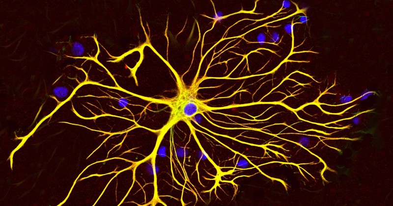
This type of glial cell was described by Ramón y Cajal. During his observations of neurons, he noticed that close to the neurons there were other cells, stellate in shape; hence its name. It is located in the CNS and along the optic nerve, and is possibly one of the glia that performs a greater number of functions.. Its size is two to ten times larger than that of a neuron, and it has very different functions
Blood-brain barrier
Blood does not flow directly into the CNS. This system is protected by the blood-brain barrier (BBB), a highly selective permeable membrane. Astrocytes actively participate in it, being in charge of filtering what can pass to the other side and what cannot.. Mainly, they allow the entry of oxygen and glucose, in order to feed the neurons.
But what happens if this barrier is damaged? In addition to the problems generated by the immune system, groups of astrocytes move to the damaged area and join together to form a provisional barrier and stop the hemorrhage.
Astrocytes have the capacity to synthesize a fibrous protein known as GFAP, with which they gain robustness, in addition to secreting another protein that allows them to gain impermeability. In parallel, the astrocytes secrete neurotrophs to stimulate regeneration in the area..
Recharging of the potassium battery
Another described function of astrocytes is their activity in maintaining the action potential. When a neuron generates an electrical impulse, it collects sodium ions (Na+) to become more positive to the outside. This process by which the electrical charges outside and inside the neurons are manipulated produces a state known as depolarization, which gives rise to electrical impulses that travel through the neuron until they end up in the synaptic space. During their journey, the cellular medium always seeks equilibrium in the electrical charge, so it loses potassium ions (K+) on this occasionto equalize with the extracellular medium.
If this were always the case, in the end a saturation of potassium ions would be generated on the outside, which would mean that these ions would stop leaving the neuron, and this would result in the inability to generate the electrical impulse. This is where the astrocytes come into play, which absorb these ions inside the neuron. absorb these ions into their interior to clean the extracellular space and allow more potassium ions to continue to be secreted.. Astrocytes have no problem with charging, since they do not communicate by electrical impulses.
4. Microglia
The last of the four most important forms of neuroglia is the microglia.. It was discovered before oligodendrocytes, but was thought to come from Blood vessels. It occupies 5 to 20 percent of the CNS glia population, and its importance is based on the fact that it is the most important form of neuroglia.Its importance is based on the fact that it is the basis of the brain's immune system. Because it is protected by the blood-brain barrier, cells are not allowed to pass freely, including those of the immune system. Therefore, the brain needs its own immune system, the brain needs its own defense system, and this is formed by this type of glia..
The immune system of the CNS
This glia cell is highly mobile, which allows it to react quickly to any problem it encounters in the CNS. Microglia have the ability to devour damaged cells, bacteria and viruses, as well as to release a number of chemical agents with which to fight off invaders. But but the use of these elements can cause collateral damage, as it is also toxic to neurons.. Therefore, after the confrontation they have to produce, as do astrocytes, neurotrophs to facilitate the regeneration of the affected area.
Earlier I spoke of damage to the BBB, a problem generated in part by the side effects of microglia when leukocytes cross the BBB and pass into the brain. The interior of the CNS is a new world for these cells, and they react to everything as unknown as if it were a threat, generating an immune response against it. The microglia initiate the defense, provoking what we could say a "civil war", which generates a lot of damage to the neurons.which generates a lot of damage to the neurons.
Communication between glia and neurons
As you have seen, glia cells perform a wide variety of tasks. But one section that has not been clear is whether neurons and neuroglia communicate with each other. Early researchers already perceived that glia, unlike neurons, do not generate electrical impulses. But this changed when Stephen J. Smith saw how they communicate, both with each other and with neurons..
Smith had the intuition that neuroglia use calcium ion (Ca2+) to transmit information, since this element is the most widely used by cells in general. In a way, he and his colleagues jumped into the pool with this belief (after all, the "popularity" of an ion does not tell us much about its specific functions either), but they were right.
These researchers designed an experiment that consisted of a culture of astrocytes to which fluorescent calcium was added, which allows by fluorescence microscopy to see their position. In addition, a very common neurotransmitter, glutamate, was added to the medium. The result was not long in coming. For ten minutes they could see how the fluorescence entered the astrocytes and traveled between the cells as if it were a wave.. With this experiment they demonstrated that the glia communicate with each other and with the neuron, since without the neurotransmitter the wave is not initiated.
The latest known about glial cells
More recent research has shown that glia detect all kinds of neurotransmitters. Moreover, both astrocytes and microglia have the capacity to manufacture and release neurotransmitters (although these elements are called gliotransmitters because they originate from glia), thus influencing the synapses of neurons.
A current field of study is to see to what extent to what extent glial cells influence the general functioning of the brain and complex mental processes, such as learning, memory and memorysuch as learning, memory or sleep.
(Updated at Apr 13 / 2024)