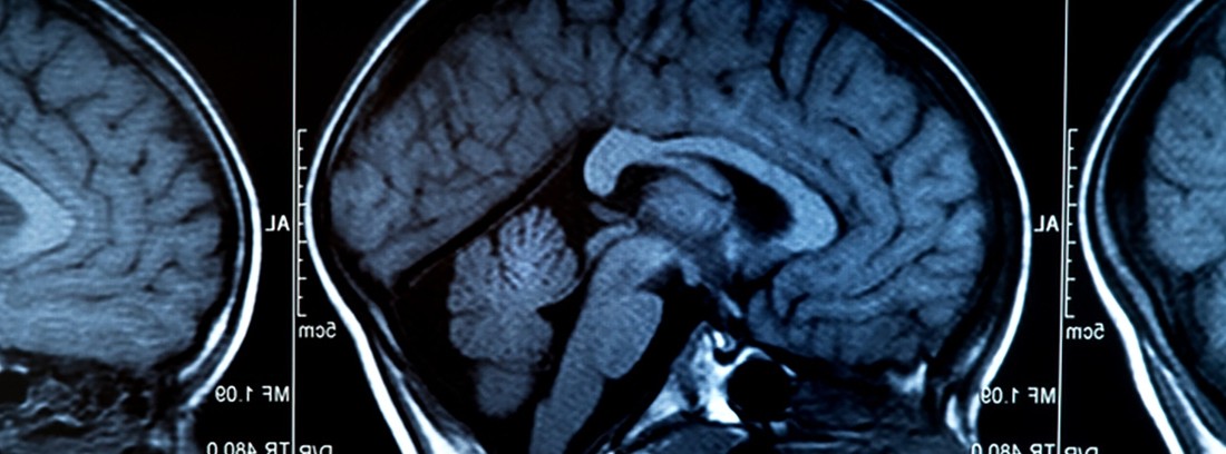
Intracranial hemorrhage

It is the breakage of a vessel inside the skull. They are classified into:
- Intraparenchymal hemorrhages or cerebral hematomas: due to rupture of the vessels that pass through the interior of the brain
- Subarachnoid hemorrhage: bleeding into the subarachnoid space where cerebrospinal fluid (CSF) normally circulates.
Non-traumatic brain hematomas account for approximately 4-14% of the causes of stroke. They have a high mortality rate, approaching 40%. The greater the bleeding, the worse the prognosis. They have a functional recovery greater than in cerebral infarction.
Subarachnoid hemorrhage constitutes 5-10% of cerebrovascular accidents. It occurs more frequently in women, between the ages of 40 and 65, with an incidence of 10 per 10,000 inhabitants. It has a high mortality, especially during the first two weeks following the acute episode, in which new bleeding is the main cause of death. Mortality is also linked to the extent of bleeding.
How is it produced?
Brain bruising occurs mainly due to high blood pressure. In it, the fact of being a chronic disease causes small dilatations to form in the cerebral arteries that at a certain moment rupture causing hemorrhage. Other causes of cerebral hemorrhage are: vascular malformations (angiomas, aneurysms), bleeding tumors, coagulation disorders, head trauma, drug abuse (cocaine, amphetamines) and arterial disorders (amioloid angiopathy).
In up to a third of patients the cause of the bleeding is not found.
In subarachnoid hemorrhage, the main cause is a ruptured aneurysm (30-60%). Aneurysms are dilatations of the vessels that make them more susceptible to rupture. A smaller percentage is produced by arteriovenous malformations and another group by alterations in coagulation, inflammatory diseases of the vessels (vasculitis), bleeding tumors and infections of the central nervous system. In young patients, the most common cause is arteriovenous malformations, in adults, aneurysms, and in the elderly, other causes.
Another group of hematomas are subdural hematomas (hematomas between the dura mater and the arachnoid) and epidural hematomas (bleeding between the dura mater and the bone), the most common cause of which is head trauma.
Symptoms
Brain hematomas usually have nonspecific symptoms such as headache (in half of the cases), nausea and vomiting until the onset of neurological deficit secondary to the location and extent of the hemorrhage. These nonspecific symptoms are what can differentiate, in the face of a sudden neurological deficit, a hemorrhage from a cerebral infarction, since clinically the neurological deficit makes them indistinguishable.
Subarachnoid hemorrhage presents as a very severe headache, nausea and vomiting, and a stiff neck, suddenly. Up to half of patients have a decreased level of consciousness that can progress to a coma.
Epidural hematomas present with an alteration in the level of consciousness, generally after a period of lucidity after a previous head injury. Acute subdural hematomas appear as a rapid and progressive deterioration towards coma, and chronic ones, with behavioral alterations, headache and tenderness to palpation in the location of the hematoma.
Diagnosis
The main imaging test that detects 100% of intraparenchymal hemorrhages and 95% of subarachnoid hemorrhages is the head CT scan. Likewise, secondary causes of bleeding, such as arteriovenous malformations, can be ruled out, the diagnosis of which must be quickly evaluated due to its potential surgical treatment.
In cases of doubtful CT, or no possibility of performing any imaging test, or in the presence of compatible symptoms without an observable lesion by cranial CT, a lumbar puncture is indicated, showing hemorrhagic CSF in cases of subarachnoid hemorrhage.
The physical examination of the patient shows, in addition to the neurological deficit according to the extent and location of the hemorrhage, in the fundus, an edematous papilla due to the intracranial pressure of the hemorrhage.
blood tests are aimed at looking for secondary causes of bleeding, especially signs of infection, liver or blood clotting disorders.
Treatment
In most brain hematomas, conservative treatment is carried out, that is, medical, and cases of surgical treatment are reserved for medium-sized hematomas in which there is progressive clinical worsening, with an accessible location and without the patient being in a coma
In subarachnoid hemorrhages, surgical treatment will be used early in cases of an acceptable level of consciousness, while in stuporous patients, surgery will be done delayed. In all cases, supportive treatment will be used, such as bed rest, sedative drugs, antitussives and that prevent constipation, good fluid intake through serums and later together with a normal diet, strict control of blood pressure, blood glucose and fever levels.
Secondary bleeding cases have their own treatment.
(Updated at Apr 14 / 2024)