Intracranial hypertension

It is the increase in pressure in the cranial cavity. The cranial cavity is made up of the brain parenchyma, the cerebrospinal fluid (CSF) and the vascular component (arteries and veins). When there is an increase in pressure of any of these components, as the skull is a closed and inexpansible cavity, an increase in intracranial pressure will occur with the appearance of a series of characteristic symptoms.
How is it produced?
The most frequent causes of intracranial hypertension are:
- Head injury: most common cause in childhood and adults
- Hydrocephalus: increased CSF
- Infectious Diseases: Abscesses, Meningitis, Empyema
- Brain tumors
- Metabolic disorders: Hyponatremia
- Vascular disorders: subarachnoid hemorrhage, cerebral infarction
Symptoms
The characteristic triad of intracranial hypertension is:
- Progressive chronic headache that does not improve with usual treatment, is throbbing in characteristics, and worsens in the morning
- Vomiting "shotgun", not preceded by nausea and predominantly morning
- Papilledema (papilledema): In up to half of patients with intracranial hypertension. Assessable by fundus examination
Other symptoms and signs that may also appear are: diplopia (double vision), hypertension, bradycardia, respiratory distress, impaired consciousness that can progress to coma and finally death.
Diagnosis
The diagnosis must be made quickly, since intracranial hypertension is considered a medical emergency, and it is necessary to know the cause of it as soon as possible to initiate the appropriate treatment and avoid further complications. For them, a good medical history should be taken followed by a complete physical examination.
Recent onset symptoms such as headache, vomiting, visual disturbances, changes in consciousness should be correctly reflected in the medical history, as well as finding out possible previous head trauma, as it is the most common cause of intracranial hypertension.
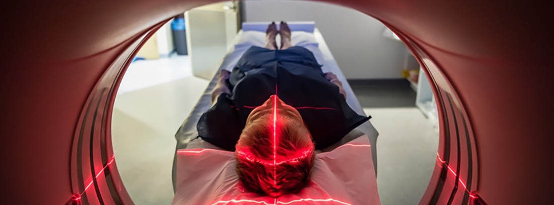
As complementary examination methods, the most useful are brain CT, brain MRI, cranial radiography, and cerebral angiography.
Treatment
In the management of intracranial hypertension, the cause of it must first be investigated, then using one treatment or another, medical or surgical. General measures are used such as stimulating ventilation, raising the head to 30-45º, ensuring a good supply of oxygen and maintaining a good balance between body fluids. Mannitol is an osmotic diuretic that is used to lower intracranial pressure. Glucorticoids are used in cases of edema associated with brain masses such as tumors.
In cases of an intracranial mass that occupies space and increases intracranial volume, as occurs in hematomas, brain tumors, cerebral edema and cerebral infarction, the treatment of choice is surgical through surgical decompression.
Family and Community Medicine Specialist
(Updated at Apr 14 / 2024)