Kidney colic: everything you need to know

The kidney stones or kidney stone It is a mineral formation that occurs inside the urinary tract due to the deposit of mineral crystals that grow to form what is known as a stone. This stone can grow in size and at any given time obstruct the passage of urine from the kidney until its expulsion through the urethra. This obstruction causes a dilation of the urinary tract that causes very intense pain that it is known as renal colic.
How is it produced?
The kidney is responsible for filtering many substances, including mineral salts. The greater the quantity of these substances in the blood plasma that the kidney clears, the greater the presence in the urine. Urine has a limited dilution capacity, that is, it can contain diluted certain substances up to a certain limit. At the moment when these substances are very abundant, the acid component of the urine can no longer dilute them further and that is when the salts precipitate and appear in a solid state. in the form of crystals in the urine. As the number of crystals increases, they join together and form an aggregate that increases in size, which is known as renal lithiasis.
Most of these crystals are made up of calcium salts, approximately 70%, especially calcium oxalate, but also calcium phosphate. Less frequent are uric acid lithiasis, approximately 10%, those of infectious origin, formed by struvite, and cystine, an amino acid that accumulates in patients suffering from cystinuria, an autosomal recessive hereditary disorder characterized by the loss in excess of this amino acid in the urine.
There are situations that can predispose to the appearance of kidney stones and, consequently, kidney colic. The states of bone turnover that promote an increase in calcium in the blood, such as some tumors, or immobilization, favor an increase in calcium in the blood and consequently in the urine. Increasing the calcium or vitamin D intake and the use of some drugs such as lithium or thiazides can also favor the formation of calcium stones.
A diet rich in protein can predispose to a increased uric acid, as well as severe muscle injuries or chemotherapy treatment.
The repeat infections they can favor the appearance of struvite stones. Likewise, any foreign body in the urinary tract, such as a catheter or urinary catheter, can cause salts to precipitate around it and form a stone.
It goes without saying that a factor that clearly influences the stone formation is that the salts that are eliminated in the urine do not have enough water to be diluted, so that a poor daily water intake will favor the appearance of kidney stones.
Symptoms
Small kidney stones are usually asymptomatic. Now, when the same flow of urine drags them and they obstruct the urinary tract, they cause a dilation of the same that causes a picture of significant pain, renal colic.
If the lithiasis is small, it can travel the entire urinary tract without giving symptoms until it reaches the urethra, where the passage is narrower and can already give symptoms, such as a feeling uncomfortable when urinating (dysuria), pain when urinating (stranguria) and from injury to the urethral wall cause bleeding (hematuria)
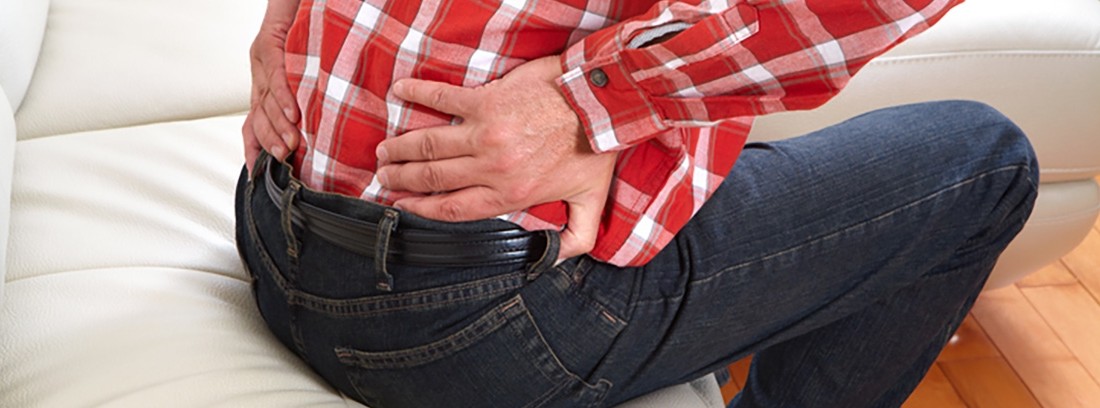
Lithiasis that obstructs the ureter will generally cause localized pain at the level of the renal fossa on the same side, that is, in the area of the back where the kidney whose ureter is obstructed is located. It's about a Intense pain and sometimes difficult to handle. It is located in this area but radiates following the path of the ureter down the side towards the lower abdomen and genitals.
It is a pain that does not subside with rest, that does not change with posture and that is always experienced, with greater or lesser intensity, but without yielding at any time. It is usually accompanied by general manifestations, such as dizziness, nausea, and even vomiting. If a fever appears, it should be suspected that the situation has been complicated by one.
When the lithiasis reaches the bladder, a temporary relief of pain usually appears, since the ureter obstruction, but irritating the bladder gives symptoms of cystitis, such as dysuria, frequency, and occasionally hematuria.
Diagnosis
The diagnosis of renal colic will be based on the symptoms that the patient presents and the physical exploration. Tapping the area of the kidney that is affected by the obstruction will produce severe sharp pain.
A urine and blood test will be performed. The urine analysis will check if there is hematuria or if there is. Due to the inflammation it is normal that there may be leukocytes but not abundant. In case of infection, a greater presence of leukocytes as well as bacteria in the urine will be seen. The blood test will allow assessing kidney function, since if there is impairment of kidney function due to an obstruction, sometimes it can lead to states of acute renal failure, the kidney will need to be unblocked immediately. It also allows assessing high levels of calcium and uric acid.
Lithiasis can be seen mostly on a plain abdominal X-ray due to its calcium composition. However, those of uric acid or other compositions will not be appreciated. The ultrasound will allow to see lithiasis of any salt, but it does not allow exploration of the middle area of the ureter. In cases of recurrent colic, it is advisable to perform an intravenous ureterography, which allows to see the route of the urinary tract from the kidney to the urethra, or a computerized axial tomography (CT).
Treatment
The vast majority of lithiasis they expel themselves, so that you only have to treat the renal colic that they can produce. Treatment will be based on pain control with pain relievers and anti-inflammatories strong, such as ibuprofen, metamizole, or diclofenac. If oral drugs are not tolerated due to vomiting that is not controlled with antiemetics or pain that does not subside, drugs should be administered intramuscularly or intravenously.
At the time of acute pain, hydration should not be forced, as by increasing the supply of water, the kidney produces more urine, which, when passing to the ureter, dilates it more and, therefore, increases pain. The hydration should be moderate and once the pain has been controlled, increase it so that the urine can carry away the lithiasis.
In the event of impaired kidney function secondary to obstruction urinary tract must be cleared urgently. The best way is to try it by placing an endoscopic catheter that overcomes the lithiasis and allows urine to flow from the kidney to the outside. If it cannot be opened endoscopically, a nephrostomy will be placed, that is, a catheter placed through the skin and guided by ultrasound until it reaches the renal pelvis so that the urine goes directly to the outside.
In cases of very large stones that cannot be expelled on their own, there is the option of fragment them into tiny pieces using ultrasound so that they can then be expelled naturally: this is known as extracorporeal shock wave lithotripsy (ESWL). In case of not being able to perform or of complicated calculations, we will proceed to the surgical extraction, either by endoscopic route, through the urine conduit, fragmenting them, or by open surgery.
Precautionary measures
The best way to prevent the appearance of lithiasis is through a correct hydration, with a daily water intake of at least 1.5-2 liters. Likewise, it is advisable to limit the intake of large meals rich in protein, especially if there are levels of elevated uric acid, as well as reducing its levels by means of allopurinol, always, of course, under a medical prescription.
(Updated at Apr 14 / 2024)