Rhinoscopy: A Closer Look Inside the Nasal Passages

Definition
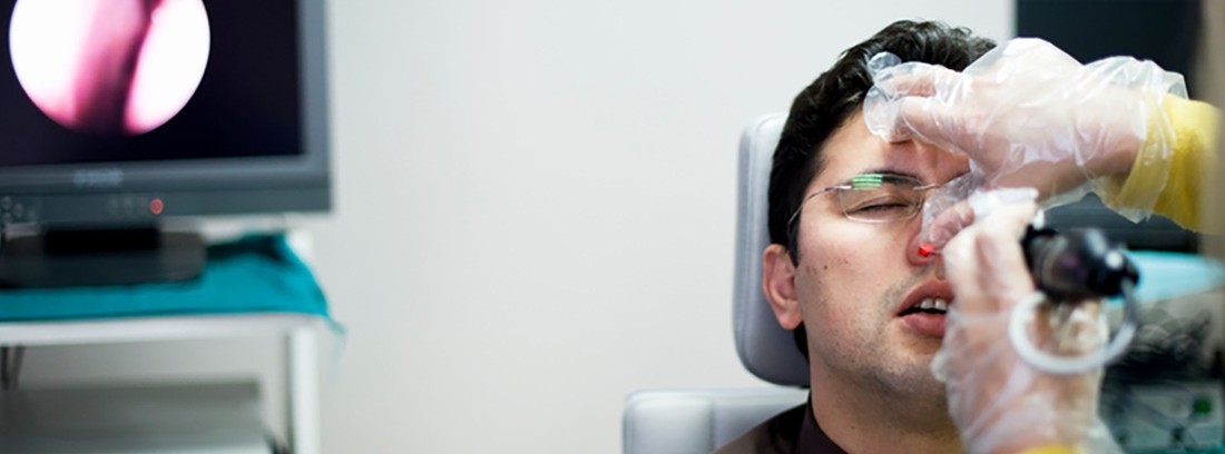
Rhinoscopy consists of visualizing the inside of the nostrils with the help of a rhinoscope or a speculum, a frontal mirror, and a light source. The rhinoscope is a widely used diagnostic instrument in the field of Otolaryngology that allows the nasal wing (nostril) to be separated from the nasal septum, thus increasing the field of vision inside the nostril.
Depending on the technique used, rhinoscopy can be classified into:
- Anterior rhinoscopy: consists of the visualization of the elements that make up the anterior portion of the nasal fossa (mucosa, nasal vestibule, inferior turbinate and sometimes middle, roof of the nostrils and choanas). For its realization a rhinoscope and an external light source are used.
- Posterior rhinoscopy: consists of the visualization of the elements that make up the posterior portion of the nostrils (superior turbinate, middle turbinate, tail of the inferior turbinate and vomer). For its realization, a speculum, a frontal mirror and an external light source are used.
Alternative names
Nasoscopy.
How is the study done?
The rhinoscopy is performed by the ENT specialist in his own practice.
In the anterior rhinoscopy, the patient remains seated on an armchair, with his head straight, his mouth closed, and breathing calmly through his nose. The doctor turns on a lamp located in front of him and a mirror is placed on his forehead into which he will project the light from the lamp and direct it towards the patient's nose in order to perform the visualization; You will place the rhinoscope inside one of the nostrils, opening it little by little to separate the nasal wing from the rest of the nasal elements and thus proceed to the visual inspection of the inside of the nostril. You will repeat the process with the contralateral nostril.
In the posterior rhinoscopy, the patient remains seated on an armchair, with the head straight and slightly forward, the mouth open, the tongue relaxed inside, and breathing calmly through the nose and mouth. The doctor turns on a lamp located in front of him and a mirror is placed on his forehead into which he will project the light from the lamp and direct it towards the patient's mouth in order to perform the visualization; He will place a speculum in your oropharynx and will proceed to a visual inspection of the posterior portion of both nostrils.
The study usually lasts between 5-10 minutes.
Preparation for the study
Rhinoscopy does not require prior preparation by the patient, although it may be advisable to clean both nostrils by using saline solutions for nasal application.
Anterior rhinoscopy may require the use of a vasoconstrictor.
Posterior rhinoscopy may require the use of a topical anesthetic during the examination if the patient is nauseous.
What does it feel like before and after the study?
Rhinoscopy is painless for the patient.
Study risks
During the study may appear:
- allergy to the drug (anesthetic and / or vasoconstrictor) used during the examination.
- Mild oropharyngeal irritation
Study contraindications
Rhinoscopy has no contraindications for its performance.
What is the study done for?
Rhinoscopy study is a very simple and safe technique, commonly used in the field of Otorhinolaryngology as it offers very useful information for the doctor as it allows the identification of possible causes of symptoms such as snoring, nasal bleeding, difficulty in passing air and infections. recurrent nasal, oropharyngeal, or otologic; among many others.
It allows to detect and / or diagnose inflammatory, infectious and / or tumor type processes that affect the interior of the nasal fossa such as rhinitis, turbinate hypertrophy, adenoid hypertrophy, or the presence of polyps, among many other processes.
It allows to identify and extract foreign bodies located in the nostril.
(Updated at Apr 13 / 2024)