Scintigraphy

Alternative names
Tracking.
Definition
Scintigraphy consists of obtaining scintigraphic images of the anatomical area to be studied using a gamma-ray emitting source (radiopharmaceutical or probe), a gamma-ray capturing source (gamma camera) and a computer.
The basis of operation of the scintigraphy is that after the administration of a specific type of radiopharmaceutical that will have been previously selected based on the tissue that we want to analyze, it will accumulate in greater or lesser concentration on said tissue and will begin to emit gamma radiation of greater or lesser intensity depending on the amount of radiopharmaceutical accumulated.
The different gamma radiation emitted will be captured by the gamma camera, giving rise to different scintigraphic images that will be sent to a computer for their definition and later study.
Depending on the anatomical area that you want to study, the scintigraphy can have different names, the most common are:
- thyroid scan
How is the study done?
The scan is performed in the radiology room of the medical center or hospital by a radiology technician. The patient must undress the anatomical area under study and, if necessary, they will be provided with a gown to cover themselves; at the same time you should remove your personal items, especially jewelry and metal objects that can interfere with radiological images.
Initially, the radiopharmaceutical will be administered, generally intravenously through a vein in the arm or hand; although in some studies it may be administered by inhalation or orally. The patient must wait in a room for about 60 minutes for the radiopharmaceutical to be fully distributed throughout the body, avoiding speech and movement as far as possible.
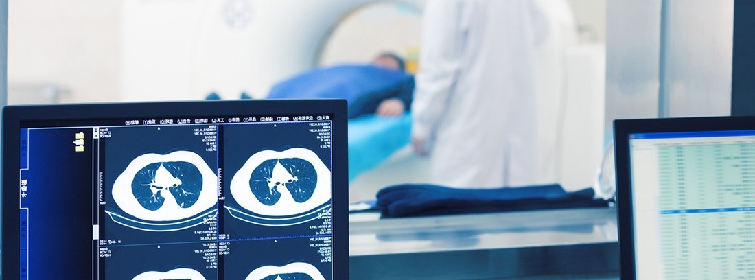
Once the radiopharmaceutical has been distributed through the tissues to be studied, scintigraphic images will be taken for which the patient will lie motionless on a stretcher while one or two gamma cameras move above and / or below the studied area.
Sometimes the gamma camera is located inside a scanner (like a tube), in these cases, it will be the stretcher on which the patient is located that moves slowly into the scanner. The images obtained by the scintigraphy will be sent to a computer for their definition and later study.
The duration of the exam will depend on the anatomical area to be studied and the amount of images necessary to complete the study, generally it usually takes 30 minutes.
Preparation for the study.
The scintigraphy does not require prior preparation but the signing of an informed consent by the patient will be requested. The patient must undress the anatomical area under study and remove their personal items, especially jewelry and metal objects.
What does it feel like during and after the study?
The scintigraphy is painless for the patient except for the discomfort of the administration of the radiopharmaceutical. The patient must remain motionless while the study is performed. In some cases, an allergic reaction to the radiopharmaceutical may occur, in the case of a skin rash, itching or respiratory distress during the study, it should be indicated to the radiology technician immediately.
Radiology rooms must be kept at a certain temperature, generally below the temperature of other rooms. The patient can lead a normal life once the study is finished except for specific indications from the doctor or the technician who has carried out the study.
Study risks.
The scan does not pose a health risk. The type of radiopharmaceutical as well as the dose used follows strict safety controls and in general the benefit obtained outweighs the minimal risks of the radiation itself. The amount of radiation emitted by the radiopharmaceutical is minimal and has a half-life of a few hours. The elimination of the radiopharmaceutical is carried out by the kidney and / or fecal route in the course of the next hours or days.
An allergic reaction to the radiopharmaceutical may occur. Severe anaphylactic reaction is rare.
The embryo, fetus, and children are more susceptible to radiation, so in these cases unnecessary studies should be avoided. Women who are pregnant or suspected of being pregnant (including those with an IUD) should avoid the study as much as possible and should tell the radiology technician that they are pregnant before having a scan.
Contraindications to the study
The patient should consult with his doctor before carrying out the study in case of:
- Pregnancy and breastfeeding.
- Kidney or liver failure
- Perform some type of treatment
- Radiopharmaceutical allergy
- You have carried out a previous study two months before.
What is the study done for?
Scintigraphy is a relatively simple and safe test, widely used in the field of Medicine as it provides very valuable information for the doctor on functional and / or metabolic alterations of the internal organs.
It allows the study and monitoring of multiple conditions such as thyroid function, brain alterations (inflammations, infections, tumor lesions, dementia ...); cardiological alterations (percussion deficit, myocardial ischemia…); tumor and / or metastatic lesions…, among many others.
Family and Community Medicine Specialist
(Updated at Apr 14 / 2024)